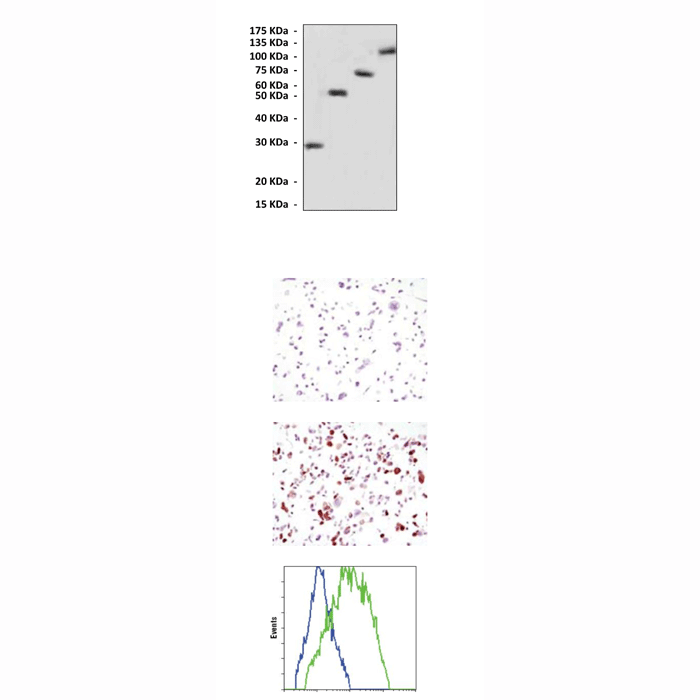
Top: Western Blot detection of various GFP-tagged proteins in transfected 293 cell lysates using GFP Antibody. Middle, upper: This antibody stains HCC827 cell lysates transfected with GFP-expression vector or mock vector. Middle, lower: This antibody stains HCC827 cell lysates transfected with mock vector in ICC testing. Bottom: This antibody specifically react
...Anti-GFP: Mouse Green Fluorescent Protein Antibody
Mouse Green Fluorescent Protein Antibody: Mouse Green Fluorescent Protein Antibody
Size: 100 ul
Price: $354.00
Description
BACKGROUND The ability to visualize organelle localization and dynamics is very useful in studying cellular physiological events. Until recently, this has been accomplished using a variety of staining methods. However, staining can give inaccurate information due to nonspecific staining, diffusion of the stain or through toxic effects. The ability to target green fluorescent protein (GFP) to various organelles allows for specific labeling of organelles in vivo. GFP is a jellyfish Aequorea victoria protein composed of 238 amino acid residues (26.9kDa).1 It exhibits bright green fluorescence when exposed to blue light. When introduced into eukaryotic cells, GFP gene produces a fluorescent protein without the need for exogenous substrates or cofactors. GFP fluorescence is stable under fixation conditions and suitable for a variety of applications. The GFP-tagged genes now provide a powerful and more direct visual demonstration of the localization of the fusion protein.2 GFP can also be used for other studies including protein-protein interaction by fluorescence energy transfer (FRET) assay.
REFERENCES
1. Zimmer, M.: Chem. Rev. 102:759-82, 2002
2. Pollok, B.A. & Heim, R.: Trend in Cell Biol. 9:57-60, 1999
2. Pollok, B.A. & Heim, R.: Trend in Cell Biol. 9:57-60, 1999
Products are for research use only. They are not intended for human, animal, or diagnostic applications.
Details
| Cat.No.: | CP10114 |
| Antigen: | Purified recombinant GFP protein expressed in E. coli. |
| Isotype: | Mouse IgG |
| Species & predicted species cross- reactivity ( ): | Human |
| Applications & Suggested starting dilutions:* | WB 1:1000 IP n/d IHC n/d ICC 1:200 FACS 1:200 |
| Predicted Molecular Weight of protein: | 27 kDa |
| Specificity/Sensitivity: | Detects GFP proteins without cross-reactivity with other fluorescent proteins. |
| Storage: | Store at -20°C, 4°C for frequent use. Avoid repeated freeze-thaw cycles. |
*Optimal working dilutions must be determined by end user.