Urogenital
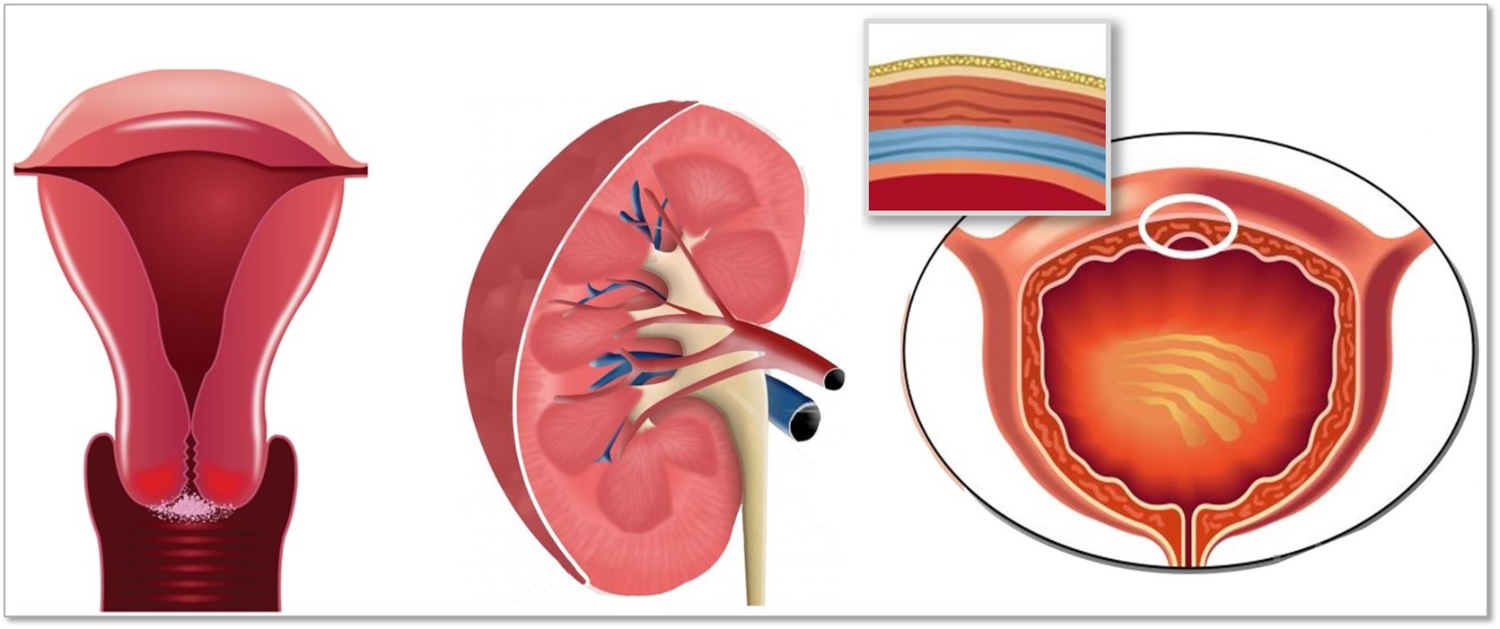
The urogenital tract is a complex, anatomically contiguous system comprising the urinary organs responsible for fluid filtration and excretion, alongside the reproductive structures required for gametogenesis and gestation. Structurally, the tract is characterized by stark regional specialization, transitioning from the high-pressure, highly permeable vascular filters of the kidney to the specialized barrier epithelia of the lower urinary tract and the cyclically remodeling mucosal linings of the reproductive organs.
Because accessing healthy, intact human urogenital tissue poses significant clinical and ethical challenges, in vitro models mapping these niches rely on a combination of human primary cells and established mammalian large-animal models (such as bovine systems). These platforms serve as crucial tools for understanding mucosal immunity, modeling barrier breakdown in urinary tract infections (UTIs), investigating endocrine-driven tissue remodeling, evaluating nephrotoxicity pipelines, and identifying therapeutic targets for urological and reproductive pathologies.
The urogenital system features a diverse array of epithelial cell types, each adapted to distinct physiological barriers and secretory demands:
The Lower Urinary Tract Barrier Niche: The bladder and urethra are lined by specialized epithelial barriers engineered to withstand mechanical stretching and resist the chemical stress of toxic metabolic waste. Human Bladder Epithelial Cells (HBlEpC)—which include transitional epithelial cells, or urothelium—form a highly impermeable barrier under normal physiological conditions to prevent the reabsorption of excreted urine. However, this barrier capacity is context-dependent, as exposure to toxins, localized ischemia, or pathological inflammation can significantly alter permeability. Further down the tract, Human Urethra Epithelial Cells (HUEpC) line the urogenital exit pathway, serving as a primary defense line against ascending pathogens. Both cell types are utilized to study the pathogenesis of uropathogenic Escherichia coli (UPEC) colonization and intracellular bacterial community (IBC) formation.
The Glandular Reproductive and Secretory Niches: Within the male reproductive tract, Human Prostate Epithelial Cells (HPrEpC) form the secretory lining of the prostate gland. These cells are highly sensitive to androgens and are critical for modeling normal prostatic exocrine function, benign prostatic hyperplasia (BPH), and the early microenvironmental changes that drive prostatic oncogenesis. In the female reproductive tract, animal models like Bovine Endometrial Epithelial Cells (BEnEpC) provide an accessible platform to investigate the uterine mucosal environment, such as studying maternal-embryo crosstalk, implantation mechanics, and the inflammatory pathways associated with endometritis. However, investigators must account for species-specific caveats—including differences in hormonal cycles, receptor expression profiles, and innate immune responses—that can limit direct extrapolation from bovine models to human reproductive physiology.
The Specialized Renal Nephron Niches: Within the functional units of the kidney, epithelial cells execute precise filtration and reabsorption duties. Human Renal Glomerular Epithelial Cells (HRGEpC), also known as visceral epithelial cells or podocytes, possess complex interdigitating foot processes that form the size-selective filtration barrier of the glomerulus. HRGEpC are highly sensitive to mechanical and metabolic stress, making them vital for studying podocyte injury in diabetic nephropathy. Downstream of the glomerulus, Human Renal Proximal Tubular Epithelial Cells (HRPTEpC) line the proximal convoluted tubule, executing the bulk of solute reabsorption and active transport. Because HRPTEpC are highly enriched with transporter networks, they are a primary target for drug-induced kidney injury (DIKI) and serve as a core model for preclinical nephrotoxicity screenings.
The structural and filtration capacities of the upper urinary tract depend heavily on specialized vascular beds that manage systemic blood flow and structural tone. Bovine macrovascular models are frequently leveraged to study these dynamics due to their physiological scale and biochemical consistency:
The Renal Arterial Endothelium: Bovine Renal Artery Endothelial Cells (BRAEC) form the inner monolayer of the renal artery. This population is specialized to handle significant hydrostatic pressure and regulate renal blood flow via the synthesis of vasoactive factors like nitric oxide and endothelin-1. BRAEC models are valuable for investigating vascular endothelial dysfunction, macrovascular inflammation, and the cellular responses to fluid shear stress within the renal circulation.
The Renal Arterial Smooth Muscle: Surrounding the endothelial lining is the tunica media, populated by Bovine Renal Artery Smooth Muscle Cells (BRASMC). These cells control the contraction and relaxation of the vessel wall, maintaining vascular tone and blood pressure regulation via auto-regulatory mechanisms. Matching BRAEC with BRASMC in co-culture systems allows researchers to model intercellular vascular crosstalk, vascular remodeling, and the phenotypic switching of smooth muscle cells from a contractile to a proliferative synthetic state during chronic hypertension or vascular injury.
