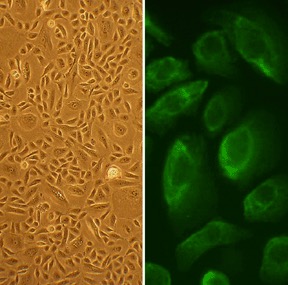
Human Bladder Epithelial Cells: HBlEpC
Human Bladder Epithelial Cells (HBlEpC) are primary epithelial cells isolated directly from the specialized lining, or urothelium, of the human urinary bladder.
Description
Human Bladder Epithelial Cells (HBlEpC), also referred to as human BDEC, are primary cells isolated directly from the specialized lining, or urothelium, of the human urinary bladder. Unlike continuous cell lines that often display altered chromosomal structures and lost differentiation capabilities, these primary bladder epithelial cells retain their physiological structural and functional characteristics in vitro. This makes them an invaluable primary cell model for studying tissue-specific cell biology.
As with other fastidious human primary cells, the behavior, lifespan, and differentiation potential of a human bladder epithelial cell depend tightly on cell culture conditions. In professional cell biologics workflows, these frozen cells are typically preserved under liquid nitrogen to maintain maximum viability. Upon thawing, they are cultured using a specialized, Human Bladder Epithelial Cell Growth Medium optimized to support healthy proliferation while suppressing fibroblastic overgrowth. Standard epithelial quality control protocols involve validating their characteristic cobblestone cell type morphology and verifying the expression of standard structural tested marker cytokeratin 18 (CK18).
In vivo, these cells are native to the mucosal layer of the urinary bladder wall. Within intact human bladder tissue, the bladder epithelium forms a highly stratified architecture organized into basal, intermediate, and specialized superficial “umbrella” cells. This stratified epithelial tissue rests upon the lamina propria — a supportive connective tissue layer containing vasculature, nerves, resident fibroblasts, and immune cells — which separates the mucosal lining from the deeper detrusor smooth muscle cell layers.
The unique structural layout of these bladder cells allows the tissue to remain highly impermeable while undergoing dramatic physical adjustments. The umbrella cells feature specialized tight junctions and asymmetric unit membranes that withstand regular stretch and contraction cycles during bladder filling and emptying.
The primary function of the bladder epithelium is to serve as a robust, specialized physical barrier that protects the underlying bladder wall and deeper tissues from urine, metabolic waste products, and potential pathogens. By maintaining strict barrier impermeability, the urothelium prevents toxic solutes and concentrated urine components from diffusing back into the deeper bladder wall layers.
Beyond acting as a passive shield, these cells actively participate in regional cell signaling and defense mechanisms. When exposed to infectious agents or potential pathogens, the epithelial cells respond by altering the transcription of specific genes, modulating cell-adhesion molecules, and secreting inflammatory cytokines to recruit immune cells to the barrier zone. However, if this barrier is chronically disrupted or exposed to environmental carcinogens, multifactorial pathobiological changes can contribute to chronic inflammation, recurring infection susceptibility, or malignant transformation.
In modern biomedical and pharmaceutical research, HBlEpC are widely recognized as a useful model system to investigate urothelial development, barrier kinetics, and oncogenesis. In cancer biology, they serve as a critical normal control in studies investigating biomarker expression, such as evaluating CD44-related variants like CD44v6 to distinguish benign tissue from urothelial carcinoma of the bladder. Researchers also utilize these primary cells to evaluate how intracellular signaling pathways adapt when the bladder tissue is exposed to uropathogenic E. coli or chemical toxins.
HBlEpC are used in advanced cell culture models, including co-culture configurations with human bladder fibroblasts or smooth muscle cells, to map the complex cellular cross-talk that drives tissue repair and remodeling following injury. By comparing data from HBlEpC alongside other specialized epithelial models — such as human nasal epithelial cells, human bronchi, and airway epithelial cells — investigators can successfully isolate organ-specific disease mechanisms from generalized epithelial responses, accelerating the development of targeted urological therapeutics.
Details
| Tissue | Normal healthy human bladder | |
|---|---|---|
| QC | No bacteria, yeast, fungi, mycoplasma, virus | |
| Bioassay | Attach, spread, proliferate in Growth Med | |
| Cryovial | 500,000 HBlEpC (2nd passage) in Basal Medium w/ 10% FBS, 10% DMSO | |
| Kit | Cryovial frozen HBlEpC (938-05a), Grwth Med (217-500), Subculture Rgnt Kit (090K) | |
| Proliferating | Shipped in Tsfr Med, psg 3, flasks or plates | |
| Doublings | At least 5 | |
| Applications | Laboratory research use only (RUO). Not for human, clinical, diagnostic or veterinary use. |
Resources
FAQs
Need More Help?
Visit our comprehensive FAQ page for detailed answers to common questions.
Need More Help?
Visit our comprehensive FAQ page for detailed answers to common questions.
Primary Cell FAQs Powered by Bioz