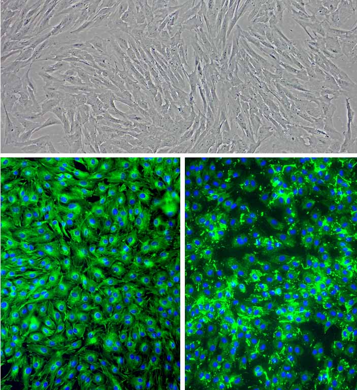
Porcine Corneal Keratocytes: PCK
Porcine Corneal Keratocytes (PCK) are specialized, neural crest-derived mesenchymal cells that populate the dense stromal layer of the porcine eye.
Description
Porcine Corneal Keratocytes (PCK), or corneal stromal keratocytes, are specialized, neural crest-derived mesenchymal cells that populate the dense stromal layer of the porcine eye. The stroma comprises the vast majority of the corneal thickness and is responsible for maintaining the structural framework and optical clarity of the tissue. This tissue architecture is composed primarily of a highly organized matrix of type I collagen, which represents the dominant stromal collagen, alongside type V collagen, which explicitly regulates fibril diameter. This structural framework is intricately organized into orthogonal lamellae alongside specialized proteoglycans to minimize light scattering.
Because of the anatomical, optical, and biochemical similarities between the pig eye and the human cornea, PCK serve as a highly relevant, translationally robust in vitro cell model in vision research. They are widely utilized to study keratocyte differentiation, structural corneal regeneration, and the pathophysiological mechanisms underpinning severe corneal blindness.
Following a mechanical or chemical injury, resident corneal stromal keratocytes exhibit a highly coordinated spatial and temporal response. Cytokines and growth factor signals released by the damaged overlying corneal epithelium enter the stroma via a combination of passive diffusion and active transport mechanisms. This triggers an immediate, localized wave of keratocyte apoptosis and cell death directly adjacent to the wound site.
Subsequently, surviving cells located more peripherally are stimulated to proliferate and transition into mobile fibroblasts (or corneal fibroblasts). If the injury breaks the epithelial basement membrane, prolonged exposure to transforming growth factor-beta (TGF-β) drives these stromal fibroblasts to further differentiate into alpha-smooth muscle actin (α-SMA)-positive myofibroblasts. While these contractile cells are necessary for rapid wound healing, their persistent activity leads to a disorganized extracellular matrix, resulting in permanent corneal opacity and structural scarring.
In the laboratory, primary Porcine Corneal Keratocytes (PCK) can be isolated from fresh tissue and successfully stored in liquid nitrogen repositories for long-term archiving. When thawed and maintained in specialized Porcine Corneal Keratocyte Growth Medium, primary PCK retain their quiescent, dendritic morphology and baseline gene expression profiles, allowing them to propagate up to 10 population doublings under recommended conditions.
However, investigators must account for culture-induced phenotypic shifts: the introduction of serum into the medium acts as an analytical switch, rapidly forcing quiescent keratocytes into a highly proliferative, fibroblastic phenotype that mirrors the active in vivo response to severe trauma.
-
Epithelial-Stromal Interactions in Corneal Wound Healing:
A major focus of ophthalmic literature, frequently highlighted in journals like Experimental Eye Research (* exp eye res *), is deciphering the paracrine loops between the porcine corneal epithelium and the underlying stroma. Researchers utilize co-culture models combining PCK with Porcine Corneal Epithelial Cells to observe how injured epithelial cells secrete specific cytokines (such as IL-1 and TNF-α) that modulate stromal cell viability. This framework is essential for mapping the early molecular events governing coordinated corneal wound healing and localized tissue remodeling.
-
Characterization of Phenotypic Markers via Immunofluorescence:
Distinguishing quiescent keratocytes from activated corneal fibroblasts or scar-forming myofibroblasts requires precise molecular indexing. Investigators routinely use immunofluorescence microscopy to track the spatial distribution and expression of definitive lineage markers. Quiescent porcine keratocytes are characterized by the high expression of aldehyde dehydrogenase (ALDH), a corneal crystallin and a robust metabolic marker highly associated with the quiescent phenotype and corneal transparency. Transition into the active proliferative phenotype is marked by a sharp downregulation of aldehyde dehydrogenase and the structural upregulation of α-SMA and fibronectin.
-
Translational Paradigms for Corneal Transplantation and Bioengineering:
Due to the global shortage of donor tissue for corneal transplantation, primary PCK provide a crucial testing matrix for bioengineered lamellar grafts and artificial matrices. Researchers evaluate how porcine keratocytes migrate into, populate, and remodel novel biomaterials. Understanding how to support functional keratocyte repopulation without triggering myofibroblast differentiation is critical for developing clear, bio-synthetic alternatives capable of restoring vision without inducing chronic graft rejection or opacity.
-
Endothelial-Stromal Homeostasis and Edema Regulation:
Proper corneal hydration is a strict requirement for transparency and is governed by a delicate fluid balance across tissue layers. Investigators deploy primary PCK alongside corneal endothelial cells to model the paracrine mechanisms that regulate stromal hydration. Because corneal endothelial cells lack a robust proliferative capacity and act as a metabolic fluid pump, understanding how stromal cytokines influence endothelial cell health helps researchers design therapeutic interventions for pathological corneal edema and endothelial decompensation.
Details
| Tissue | Normal healthy porcine cornea | |
|---|---|---|
| QC | No bacteria, yeast, fungi, mycoplasma, virus | |
| Bioassay | Attach, spread, proliferate in Growth Med | |
| Cryovial | 500,000 PCK (1st passage) frozen in Basal Medium w/ 10% FBS, 10% DMSO | |
| Kit | Cryovial frozen PCK (P632-05), Growth Medium (P6111-500), Subculture Rgnt Kit (090K) | |
| Proliferating | Shipped in Tsfr Med, psg 1, flasks or plates | |
| Doublings | At least 15 | |
| Applications | Laboratory research use only (RUO). Not for human, clinical, diagnostic or veterinary use. |
Resources
FAQs
Need More Help?
Visit our comprehensive FAQ page for detailed answers to common questions.
Need More Help?
Visit our comprehensive FAQ page for detailed answers to common questions.
Primary Cell FAQs