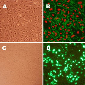
Human Aortic Endothelial Cells: HAOEC
Human Aortic Endothelial Cells (HAOEC) represent a specialized population of endothelial cells derived from the luminal surface of the aorta.
Description
Human Aortic Endothelial Cells (HAOEC) represent a specialized population of endothelial cells derived from the luminal surface of the aorta. As a primary cell type rather than an immortalized cell line, HAOEC are a widely utilized in vitro model for investigating vascular biology, maintaining critical phenotypic markers such as CD31 (PECAM-1), VE-cadherin, and von Willebrand factor (vWF). HAOEC are frequently provided as frozen cells or as a functional cell pellet, often isolated from a single donor to avoid the confounding variables of mixed genetic backgrounds. When cultured in specialized Human Endothelial Cell Growth Medium or optimized endothelial cell growth media formulas, these primary cells retain functional characteristics, including the capacity for acetylated-LDL uptake and stimulation-dependent nitric oxide production.
In the human body, HAOEC form the innermost large-vessel monolayer lining the lumen of the aorta, the main trunk of the systemic arterial network. Situated at the interface between the circulating blood and the vessel wall, this specific cell type is constantly exposed to distinct hemodynamic forces, such as fluid shear stress. While endothelial cells line all blood vessels, large-vessel endothelia exhibit pronounced regional heterogeneity when compared to populations in other vascular beds, such as the pulmonary artery, coronary artery, or adjacent microvasculature. Extensive comparative studies demonstrate that endothelia from different tissue type origins vary significantly in their baseline profiles, unique surface N-glycosylation patterns, and interactions with circulating leukocytes. This distinct spatial variation helps explain why specific arterial segments are differentially susceptible to localized vascular disease and inflammatory stimuli.
The primary function of HAOEC is to maintain vascular homeostasis by acting as a selective semipermeable barrier, regulating vascular tone, inhibiting inappropriate coagulation, and modulating immune cell transmigration. Under healthy conditions, these cells express a complex network of protective genes that manage vascular health. However, when subjected to environmental or metabolic stresses, HAOEC undergo endothelial dysfunction. For instance, increased glucose flux in diabetic configurations can trigger pro-inflammatory responses. Similarly, exposure to certain air pollutants and oxidative stressors has been shown to compromise tight junction function (such as ZO-1) or alter stress signaling pathways (like JNK and p38), though these molecular events vary depending on the specific pollutant, exposure levels, and the experimental model system. While primary macrovascular cells have a more limited inherent angiogenic capacity compared to microvascular or progenitor endothelial types, HAOEC can still exhibit angiogenic behaviors and key signaling pathways under appropriate, targeted stimuli.
In laboratory settings, HAOEC are an invaluable model for exploring the molecular mechanics of cardiovascular disease, atherosclerosis, and coronary artery disease, provided researchers account for how passage number affects the baseline phenotype. Scientists manipulate ambient conditions in cell culture to observe how advanced glycation end products, uremic toxins, or altered ambient glucose levels modify downstream gene expression and drive pathologically altered genes. Because vascular pathologies involve complex multicellular interactions, investigators frequently study HAOEC alongside a panel of primary vascular cell types (e.g., VSMCs, fibroblasts) to analyze structural cross-talk and intimal hyperplasia. Furthermore, HAOEC are utilized to evaluate the anti-inflammatory or vasodilating properties of novel therapeutic compounds, and to engineer 3D endothelialized tissues. These engineered models are critical for testing advanced biomaterial surfaces and drug-delivery coatings (such as paclitaxel or sirolimus) designed to support endothelial cell adhesion and proliferation while preventing restenosis in vascular implants.
- Demonstrate that increased glucose flux leads to endothelial dysfunction in diabetes via activating Egr1-mediated proinflammatory and prothrombotic responses
- Study apoptosis, oxidative stress and inflammation associated with atherosclerosis and demonstrate the beneficial effects of anthocyanin on endothelial cells damaged by exposure to oxidized sterols
- Demonstrate that upregulation of thioredoxin via AMPK-FOXO3 pathway protects endothelial cells from oxidative stress and may prevent cardiovascular diseases in patients with metabolic syndrome and diabetes and further elucidate the involvement of AMPK cascade in mediating beneficial cardiovascular effects of green tea
- Test anti-inflammatory and vasodilating properties of a synthetic rutaecarpine derivative
- Show that glycated albumin, associated with diabetic complications, decreases endothelial miR-146a expression which leads to increased IL-6 production, and that angiotensin protects endothelial cells by preventing miR-146a downregulation
- Demonstrate that air pollutants can directly affect ZO-1 function leading to increased endothelial permeability, inflammatory cell transmigration and initiation of atherosclerosis
- Discover the involvement of stress signaling JNK and p38 pathways in pathological suppression of thrombomodulin, a vascular protective molecule, downregulated in many thrombotic and vascular diseases
- Link uremic toxins (in particular, PAA) in patients with chronic liver disease to increased ROS production and stimulation of TNF-a in endothelial cells leading to atherosclerosis and vascular calcification
- Demonstrate that in diabetes, advanced glycation end products lead to ROS generation in endothelia via sustained NF-kB activation, contributing to progression of atherosclerosis
- Discover that CD40 ligand promotes monocyte adhesion to endothelial cells via PKCa, NF-kB and VCAM-1 signaling cascade, explaining the role of CD40L in atherogenesis
- Show that monocytes activated by endothelial cells, produce CD80 signaling that leads to allogenic immune response, indicating the need for specific therapy to prevent monocyte activation during allograft transplantation
- Identify tetraspanin CD82 as the recognition sensor responsible for rejection of xenotransplants
- Develop 3d endothelialized engineered tissues, as well as new technology based on novel material surfaces and drugs (such as paclitaxel, sirolimus, vitamin C, C6-ceramide and 17β-estradiol) to inhibit smooth muscle cell proliferation at the same time allowing endothelial cells adhesion and proliferation in order to reduce risk associated with vascular implants
Details
| Tissue | Normal healthy human aorta |
|---|---|
| QC | No bacteria, yeast, fungi, mycoplasma |
| Character | Factor VIII-related Ag, DiI-Ac-LDL uptake. S-HAOEC are select HAOEC lots that have been tested positive for VEGFR2 pathway activation following stimulation by VEGF. |
| Bioassay | Attach, spread, proliferate in Growth Med |
| Cryovial | 500,000 HAOEC (2nd passage) frozen in Basal Medium w/10% FBS, 10% DMSO |
| Kit | Cryovial frozen HAOEC (304-05a), Growth Medium (211-500), Subculture Rgnt Kit (090K) |
| Proliferating | Shipped in Tsfr Med, 3rd psg (flasks or plates) |
| Doublings | At least 16 |
| Applications | Laboratory research use only (RUO). Not for human, clinical, diagnostic or veterinary use. |
Resources
FAQs
Need More Help?
Visit our comprehensive FAQ page for detailed answers to common questions.
Need More Help?
Visit our comprehensive FAQ page for detailed answers to common questions.
Primary Cell FAQs Powered by Bioz