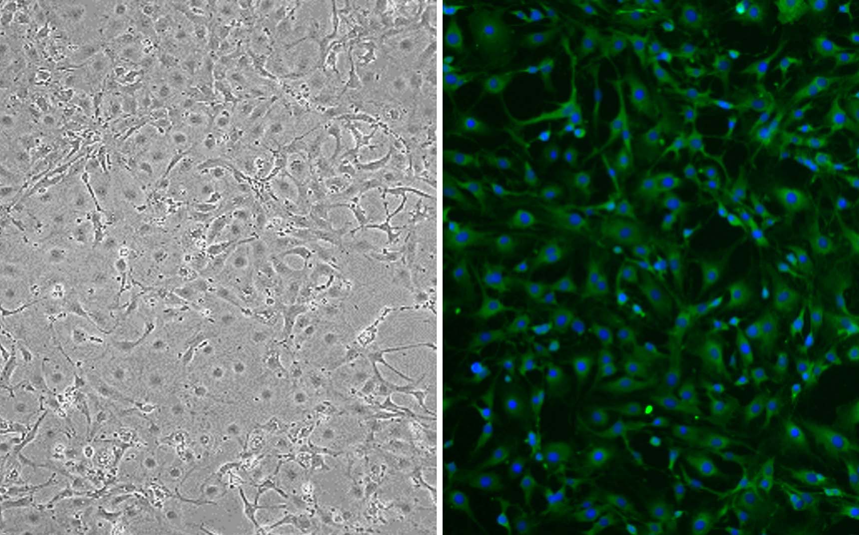
Mouse Hepatic Stellate Cells: MHSC
Mouse Hepatic Stellate Cells (MHSC) are pericyte-like, liver-resident mesenchymal cells that are isolated directly from healthy mouse liver tissue.
Description
Mouse Hepatic Stellate Cells (MHSC) are pericyte-like, liver-resident mesenchymal cells that contribute to hepatic architecture, retinoid storage/metabolism, and tissue homeostasis. These mouse primary cells are isolated directly from healthy mouse liver tissue. While their embryological origin remains a subject of ongoing debate (particularly regarding neural crest vs. mesodermal lineages), rodents express specific baseline markers such as desmin, platelet-derived growth factor receptor-beta (PDGFRβ), and glial fibrillary acidic protein (GFAP) — though investigators must note critical species differences, as GFAP expression is robust in rodent HSCs but low and highly variable in human HSCs.
In their native, non-activated state, these cells are characterized by prominent cytoplasmic lipid droplets containing retinyl esters (vitamin A) and triglycerides. These droplets exhibit a characteristic blue autofluorescence under UV/near-UV or violet/blue excitation, which is highly prone to photobleaching depending on instrumentation. Unlike immortalized lines, primary MHSCs natively maintain their physiological signaling networks. However, maintaining quiescence in vitro is technically demanding; rigid polystyrene culture plastic rapidly drives spontaneous activation. To preserve their unactivated state, investigators utilize specialized HSC culture systems featuring soft biomimetic substrates, low-serum media, and specific extracellular matrix coatings.
In the living organism, this cell type resides exclusively within the perisinusoidal space, also known as the space of Disse — a narrow subendothelial microenvironment located between the basolateral surface of hepatocytes and the fenestrated sinusoidal endothelial cells. Within this hepatic microcirculation layout, quiescent HSCs make up roughly 5–8% of the total resident cell population, though reported values vary considerably (spanning up to 15% of non-parenchymal cells) depending on the species, cell-separation method, and whether calculation baselines reflect total versus non-parenchymal counts. Here, they wrap their long cytoplasmic processes around the sinusoids, working in close paracrine proximity with liver endothelial cells, Kupffer cells, and transient immune populations.
In a healthy liver, quiescent HSCs are the liver’s principal retinoid-storage cells, storing retinyl esters and contributing to retinoid metabolism and systemic retinoid homeostasis. However, their most defining physiological feature is a profound functional reprogramming cascade known as hepatic stellate cell activation. Following acute or chronic liver injury, multiple neighboring cell types release paracrine signals (such as TGF-, PDGF, reactive oxygen species, and pro-inflammatory cytokines) that trigger indirect or direct HSC activation.
During this cascade, the cells lose their characteristic vitamin A lipid droplets, rapidly proliferate, and morphologically transition into an activated HSC phenotype. This transdifferentiation drives a marked shift in gene expression: cells upregulate alpha-smooth muscle actin (-SMA) expression—assembling -SMA–positive stress fibers that dictate a contractile, myofibroblastic phenotype—while strongly upregulating synthesis of fibrillar ECM (mainly collagens I and III). This hyper-synthetic shift alters the mechanical compliance of the liver tissue and drives sinusoidal capillarization, a destructive pathological process involving the loss of endothelial fenestrae and abnormal basement membrane deposition that disrupts normal hepatic microcirculation.
In translational gastroenterology and discovery pharmacology, MHSCs serve as widely used preclinical models that approximate aspects of human HSC biology, though investigators must account for distinct interspecies differences when translating outcomes. Researchers deploy these primary cultures to map the cell-to-cell signaling loops that drive progressive liver disease, hepatic fibrosis, and cirrhotic remodeling. By exposing cells to targeted growth factor stimulation, investigators can recapitulate the molecular triggers of HSC activation in vitro, tracking phenotypic transitions via confocal microscopy or quantifying changes in matrix-specific mRNA expression.
Furthermore, isolated HSCs function as an essential screening model to assess HSC-specific drug responses and local metabolism or toxicity. While hepatocytes remain the primary cells responsible for whole-organ systemic drug clearance, HSCs are heavily used to screen therapeutic candidates for their ability to blunt the fibrotic cascade or selectively trigger apoptosis in activated HSCs. These workflows often compare in vitro cellular responses against tissue samples from treated mice with induced liver fibrosis, evaluating whether candidate molecules can halt hepatic fibrogenesis without injuring surrounding hepatocytes or neighboring portal fibroblasts. To thoroughly evaluate in vitro activation or its attenuation by drug candidates, investigators routinely track an expanded panel of additional activation markers (such as TIMP1, Col1A1, desmin, and PDGFRβ) alongside functional assays measuring cell proliferation, contractility, and collagen secretion.
Mouse Hepatic Stellate Cells (MHSC) are one of the important cell type in the liver. Hepatic Stellate Cells play an important role in liver homeostasis, maintenance and regeneration including retinol metabolism. Activation of stellate cells in disease state leads to liver fibrosis, hepatitis B & C, fatty liver disease, diabetes and other chronic diseases. Hepatic Stellate Cells are primary cells derived from mouse liver and cultured in optimized Hepatic Stellate Cells Growth Medium. MHSCs from Cell Applications, Inc. provide an excellent model system to study many aspects of liver function, metabolism and pathophysiology. Primary culture of Hepatic Stellate Cells is suitable for many cell-based assays including toxicity, drug screening and metabolism and is a valuable tool to study liver function, physiology and liver diseases. Characterization: Positive for α-smooth muscle actin by ICC staining.
Details
| Tissue | Normal healthy mouse liver tissue | |
|---|---|---|
| QC | No bacteria, yeast, fungi, mycoplasma, virus | |
| Bioassay | Attach, spread, proliferate in Growth Med | |
| Cryovial | 500,000 MHSC (3rd passage) frozen in Basal Medium w/ 10% FBS, 10% DMSO | |
| Kit | Cryovial frozen MHSC (M782-05a), Growth Medium (M7117-500), Subcltr Rgnt Kit (090K) | |
| Proliferating | Shipped in Gr Med, 3rd psg (flasks or plates) | |
| Doublings | At least 10 | |
| Applications | Laboratory research use only (RUO). Not for human, clinical, diagnostic or veterinary use. |
Resources
FAQs
Need More Help?
Visit our comprehensive FAQ page for detailed answers to common questions.
Need More Help?
Visit our comprehensive FAQ page for detailed answers to common questions.
Primary Cell FAQs