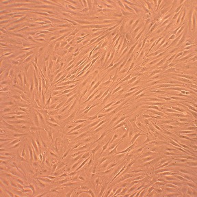
Human Fibroblast-Like Synoviocytes-Rheumatoid Arthritis: HFLS-RA. Cells that line the joints, producing synovial fluid. In RA, synovial cells express genes and erosive enzymes that contribute to joint degradation.
Human Fibroblast-Like Synoviocytes: Rheumatoid Arthritis: HFLS-RA
Human Fibroblast-Like Synoviocytes: Rheumatoid Arthritis (HFLS-RA) are a pathologically altered mesenchymal-derived cell population obtained from the hyperplastic synovial tissue of patients diagnosed with the chronic autoimmune joint disease rheumatoid arthritis.
Description
Human Fibroblast-Like Synoviocytes: Rheumatoid Arthritis (HFLS-RA) — sometimes referred to as rheumatoid arthritis synovial fibroblasts (RASFs) — are a pathologically altered mesenchymal-derived cell population obtained from the hyperplastic synovial tissue of patients diagnosed with chronic autoimmune joint disease. Unlike a transformed or continuous cell line, these human fibroblast-like synoviocyte populations are true primary cell models that inherently retain a pathologically primed “disease memory” in vitro. Structurally, these fibroblasts display an elongated, spindle-shaped morphology and function as the dominant resident cell type driving the destruction of the joint architectural matrix. To sustain their aggressively activated phenotype and prevent premature senescence ex vivo, they are maintained using a highly optimized culture system, such as an HFLS basal medium enriched with specific growth factors.
In a suffering RA patient, these specialized fibroblasts reside within the lining layer and sublining regions of the joint synovium. Under normal physiological conditions, the synovial membrane remains a thin, delicate structure, but during active RA pathogenesis, the tissue undergoes severe synovial hyperplasia, expanding into a thick, aggressive tissue mass known as a pannus. High-resolution single-cell mapping studies published in journals like Arthritis Rheumatol have revealed that the inflamed RA synovial tissue is populated by distinct FLS subsets and hyper-activated fibroblast subsets. This hyperplastic tissue mass actively expands across the joint cavity, outgrowing its nutrient supply to trigger localized angiogenesis by endothelial cells, and directly invades neighboring anatomical structures.
The primary biological function of healthy fibroblast-like synoviocytes is to maintain joint homeostasis, but in the setting of RA, the function of HFLS-RA undergoes a profound phenotypic transition. They switch from homeostatic stromal cells into aggressive, central drivers of joint destruction. They exhibit a distinct activated state characterized by tumor-like (invasive, apoptosis-resistant) behavior, which includes a dramatic increase in cell proliferation, autonomous migration, and a severe resistance to programmed cell death. This is a phenotypic analogy — HFLS-RA are not cancerous. Driven by interconnected inflammatory and signaling networks, they change their global gene expression profiles to hypersecrete pro-inflammatory cytokines and a destructive panel of matrix metalloproteinases (such as MMP-1, MMP-3, and MMP-13). This intensive secretion directly fuels cartilage destruction and marginal bone erosion. Furthermore, they function as immunological hubs, releasing chemokines that actively recruit, retain, and activate infiltrating immune cells, including macrophages, dendritic cells, and T cells.
In laboratory settings, primary HFLS-RA serve as a gold-standard human cell platform for interrogating complex chronic immunopathological mechanisms, mapping dysregulated intracellular cascades, and screening novel therapeutic modalities. Investigators must note that a donor’s specific prior medication history and clinical disease stage can directly influence the baseline ex vivo phenotype of these primary cells. Rather than operating via a single linear cascade, synoviocyte activation involves a highly complex, interconnected pathway network; while researchers frequently study the NF-B, Tpl2/MAP3K8, and p38 MAPK cascades, many other pathways — including JAK/STAT, alternative MAPKs, Wnt, Notch, and various epigenetic regulators — actively contribute to the disease state.
Because they preserve their aggressive disease phenotype ex vivo, discovery pipelines deploy HFLS-RA to screen diverse small-molecule inhibitors, natural products (like celastrol or triptolide), and targeted biologics designed to curb synoviocyte activation and promote apoptosis. Additionally, bioengineers use these cells to validate advanced non-viral gene-delivery systems and evaluate the cellular uptake of therapeutic liposomes, ensuring that emerging disease-modifying antirheumatic therapies (DMARDs) can safely interrupt the self-sustaining inflammatory response before it leads to irreversible skeletal damage.
Human Fibroblast-Like Synoviocytes: Rheumatoid Arthritis (HFLS-RA) provide an excellent cellular model for studying synoviocyte physiology in relation to development and treatment of rheumatoid arthritis. HFLS-RA from Cell Applications, Inc. have been used in research to evaluate:
- Signaling pathways implicated in the development of joint inflammation and rheumatoid arthritis
- Contribution of EBV to nonresolving rheumatoid arthritis inflammation through inducing IL-6 production by synoviocytes
- Anti-inflammatory and antirheumatic activity of various compounds, such as celastrol, triptolide, arsenic trioxide, bucillamine, therapeutic lyposomes, anti-IL-6R and anti-CD319 antibodies, inhibitors of NF-κB, methionine aminopeptidase-2, Tpl2, aryl hydrocarbon receptor, p38 MAP kinase
- Beneficial anti-inflammatory effects of low level light therapy
- Causes of metalloproteinase induction in patients with Lyme disease-associated arthritis
- Adenovirus-based siRNA delivery systems
- Causative agents, immunopathological mechanisms and signal transduction pathways leading to joint inflammation in rheumatoid arthritis
- The role of estrogen signaling in increasing inflammation
- Anti-inflammatory properties of herbal compound Sinomenine suggested for rheumatoid arthritis treatment
- Effects of extracellular matrix composition on cell attachment and migration relevant to T-cell function in inflamed tissues
- Involvement of capsid proteins of parvorvirus B19 in activating synoviocyte migration and induction of the inflammatory response leading to acute symmetrical polyarthropathy
- The role of human endogenous retroviruses (HERVs) in development of rheumatoid arthritis, and suggest that activated expression of different forms of HERV contribute to development of rheumatoid arthritis symptoms by different mechanisms
Details
| Tissue | Human synovial tissue from donor with rheumatoid arthritis |
|---|---|
| QC | No bacteria, yeast, fungi, mycoplasma, virus |
| Cryovial | 500,000 HFLS-RA frozen in Basal Medium w/ 10% FBS, 10% DMSO |
| Kit | Cryovial frozen HFLS-RA (408RA-05a), Gr Med (415-500), Subcltr Rgnt Kit (090K) |
| Proliferating | Shipped in Gr Med, 3rd psg (flasks or plates) |
| Doublings | At least 5 |
| Applications | Laboratory research use only (RUO). Not for human, clinical, diagnostic or veterinary use. |
Resources
FAQs
Need More Help?
Visit our comprehensive FAQ page for detailed answers to common questions.
Need More Help?
Visit our comprehensive FAQ page for detailed answers to common questions.
Primary Cell FAQs Powered by Bioz