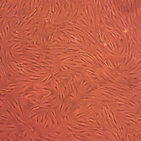
Human Fibroblast-Like Synoviocytes: Osteoarthritis: HFLS-OA
Human Fibroblast-Like Synoviocytes: Osteoarthritis (HFLS-OA) are specialized, mesenchymal-derived fibroblast cells isolated from the diseased synovial membrane of patients suffering from the degenerative joint disease osteoarthritis.
Description
Human Fibroblast-Like Synoviocytes: Osteoarthritis (HFLS-OA) — also termed osteoarthritis synovial fibroblasts or OA FLS — are specialized, mesenchymal-derived cells isolated from the diseased synovial membrane of patients suffering from degenerative joint disease. Unlike a transformed or continuous cell line, these human fibroblast-like synoviocytes are primary synovial cells that retain a pathologically primed memory of the osteoarthritic joint environment. Structurally, these fibroblasts exhibit an elongated, spindle-shaped morphology and represent a major cell type within the joint synovium. To preserve their unique biological features and prevent phenotypic drift in vitro, they are maintained using a specialized culture system, such as an HFLS basal medium supplemented with growth factors.
In the human body, these cells are localized within the synovial tissue that encapsulates diarthrodial joints, specifically concentrated within the intimal lining layer and sublining regions of the synovium. In a healthy joint, the lining layer fibroblasts regulate homeostatic joint volume, manage nutrient transport, and secrete key structural elements into the synovial fluid to lubricate the adjacent articular cartilage. However, in knee osteoarthritis and other affected joints, the local tissue architecture undergoes severe changes. High-resolution mapping studies published in journals like Arthritis Rheumatol have identified altered fibroblast subsets and distinct FLS subset populations. Driven by disease-state environments, these cells hyperproliferate, contributing to synovial tissue thickening, localized vascularization by endothelial cells, and synovial fibrosis.
The primary biological function of healthy fibroblast-like synoviocytes is to maintain joint homeostasis. However, in the chronic, low-grade inflammatory environment characteristic of osteoarthritis, the function of HFLS-OA undergoes a distinct pathological shift. Rather than remaining passive bystanders, they actively participate in joint inflammation and cartilage degradation. This multi-factorial phenotypic activation is driven by a complex combination of mechanical stress, persistent low-grade inflammation, altered local metabolic factors, cellular senescence, and continuous paracrine signaling from infiltrating immune cells. When stimulated by these joint stressors, their gene expression profiles change dramatically, shifting them into an active catabolic state where they hypersecrete matrix-degrading enzymes (such as MMP-13) and inflammatory cytokines (like IL-6 and IL-8). While they do not form the aggressive, systemic pannus typically seen in a rheumatoid arthritis (RA) patient, HFLS-OA interact closely with macrophages, dendritic cells, and T cells to perpetuate localized joint destruction.
In laboratory settings, HFLS-OA serve as a translationally rigorous human cell model for investigating osteoarthritic synoviocyte biology, understanding low-grade chronic synovial inflammation, and screening emerging chondroprotective compounds. Researchers frequently deploy HFLS-OA within comparative multi-donor cell panels alongside healthy synoviocytes and rheumatoid arthritis (HFLS-RA) cells. These comparative setups allow investigators to determine if specific mechanisms—such as the LPA₁ signaling pathway or human endogenous retrovirus (HERV) activation—are unique to an inflammatory arthritis background or represent a generalized response to chronic synovial stress.
Furthermore, following the foundational clinical frameworks established by rheumatology pioneers like Smolen J for grading joint pathologies, researchers use these cells to test advanced biomaterial scaffolds, evaluate intra-articular drug-delivery platforms, and validate novel therapeutic interventions designed to interrupt the progressive cascade of arthritis before it leads to irreversible cartilage loss.
- Possess osteogenic, chondrogenic and adipogenic potentials
- Express about half of the mesenchymal stem cell related transcription factors and molecular markers identified in a comprehensive study
- Involve LPA–EDG2 signaling in the pathogenesis of osteoarthritis via catabolic processes
- Employ parvorvirus B19 capsid proteins to activate synoviocyte migration and induction of the inflammatory response leading to acute symmetrical polyarthropathy
Details
| Tissue | Human synovial tissue from donor with osteoarthrits | |
|---|---|---|
| QC | No bacteria, yeast, fungi, mycoplasma, virus | |
| Cryovial | 500,000 HFLS-OA (2nd passage) frozen in Basal Medium w/ 10% FBS, 10% DMSO | |
| Kit | Croyvial frozen HFLS-OA (408OA-05a), Gr Med (415-500), Subcltr Rgnt Kit (090K) | |
| Proliferating | Shipped in Gr Med, 3rd psg (flasks or plates) | |
| Doublings | At least 5 | |
| Applications | Laboratory research use only (RUO). Not for human, clinical, diagnostic or veterinary use. |
Resources
FAQs
Need More Help?
Visit our comprehensive FAQ page for detailed answers to common questions.
Need More Help?
Visit our comprehensive FAQ page for detailed answers to common questions.
Primary Cell FAQs Powered by Bioz