Human Bronchial Epithelial Cells: HBEpC
Human Bronchial Epithelial Cells (HBEpC) are primary lung epithelial cells isolated from the large airways of the human respiratory tract.
Description
Human Bronchial Epithelial Cells (HBEpC) — alternatively designated as a primary human bronchial epithelial cell type or evaluated alongside normal human bronchial epithelial (NHBE) cells in literature — are primary lung epithelial cells isolated from the large airways of the human respiratory tract. Unlike a transformed continuous cell line or immortalized cell isolates, these primary airway epithelial cells retain their native diploid karyotype and exhibit more physiologically representative receptor expression than many immortalized lines. To preserve their structural integrity, prevent premature senescence, and avoid squamous dedifferentiation in vitro, they are maintained using a dedicated specialty cell culture system and specialized primary cell culture media formulations. For long-term preservation, these fastidious cells are cryopreserved and stored in liquid nitrogen vapor phase to maintain high post-thaw cell viability.
In the living organism, these cells form the continuous, pseudostratified columnar mucosal inner lining of the human trachea and the main conductive branches of the human bronchi. Situated at the interface between the host tissue and the external atmosphere, this airway epithelium rests upon a specialized basement membrane and is interspersed with distinct endogenous cell types. In the native tissue layout, multipotent basal cells act as the local progenitor pool, dividing and differentiating to replace specialized, terminally differentiated lineages.
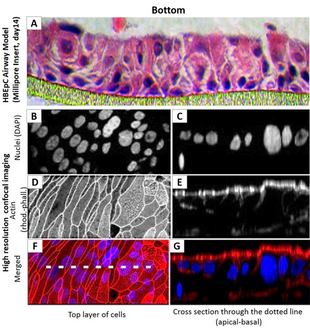
The primary biological function of this specialized cell population is to act as the frontline physical, immunological, and mechanical barrier of the respiratory tract, defending the deep lung architecture from inhaled pathogens, environmental particulate matter, and gaseous toxins. Structurally, this is achieved through strict regulation of bronchial epithelial permeability, which is maintained by complex apical tight junctions, desmosomes, and adherens junctions. Functionally, when cultured using an air liquid interface (ALI) method — commonly termed an ALI culture setup — HBEpC undergo asymmetric differentiation. They polarize into a mature, functional pseudostratified epithelium containing mucus-secreting goblet cell elements and functional ciliated cell lineages that orchestrate mucociliary clearance.
In the fields of molecular biology, functional genomics, and virology, HBEpC represent a premier human-background platform. Investigators widely deploy these primary cultures to trace host-pathogen interactions during respiratory viral infections (such as influenza, respiratory syncytial virus, and coronaviruses), mapping how cellular protection systems alter gene expression, kinase cascades, and downstream pro-inflammatory pathways. Because they display more physiologically relevant signaling and innate immune responses, they serve as an essential translational benchmark to validate or extend high-throughput screening work originally performed in less representative continuous lines. They are heavily utilized in drug discovery cell screening pipelines to evaluate novel therapeutics designed to mitigate chronic lung injury, suppress pathological apoptosis, or blunt hyper-inflammatory cascades.
Furthermore, these primary cell models are extensively integrated into respiratory disease workflows to explore the molecular pathology of chronic obstructive pulmonary disease (COPD), asthma, smoking-induced epithelial remodeling, and genetic diseases like cystic fibrosis. While normal HBEpC are not primary models for tumor transcription without additional transformation or tumor material, they are frequently integrated into specialized co-culture or tumor models to evaluate how the healthy airway epithelium interacts with surrounding malignant cells, helping researchers screen therapeutics designed to suppress tumor progression, enhance cellular protection, and accelerate normal tissue repair.
Human Bronchial Epithelial Cells (HBEpC) provide an excellent model system to study all aspects of epithelial function and disease, particularly those related to airway viral infections, as well as tissue repair mechanisms, signaling changes and potential treatments relevant to lung injuries, mechanical and oxidative stress, inflammation, pulmonary diseases and smoking. When grown on inserts and provided with the liquid/air interface, HBEpC can differentiate into a pseudostriated epithelium and serve as a more physiological 3D tissue model for in vitro studies. The HBEpC shown here were cultured (L) and immunolabeled for cytokeratin 18 (R). 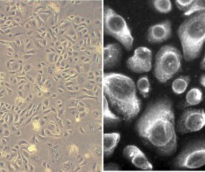
- Activation, expression and production of genes, kinases and signaling pathways by cytokines, growth factors, interleukins, binding proteins and pro-inflammatory molecules.
- Stimulation-dependent, observable changes in proliferation, bronchial epithelial permeability, crosslinking of membrane glycoproteins and cell surface adhesion molecules. Drug discovery cell screening for in vitro assay of compounds, or to extend and confirm high-throughput work done in cell lines.
- Clinical focused discoveries leveraging HBEpC include therapeutics to suppress tumor gene transcription, apoptosis, inflammation, auto-immune disease and viral infection, while enhancing cell protection, repair and lifespan.
Details
| Tissue | Human bronchial epithelium: nondiseased, Asthma, COPD, or Type 2 Diabetes | |
|---|---|---|
| QC | No bacteria, yeast, fungi, mycoplasma, virus | |
| Bioassay | Attach, spread, proliferate in Growth Med | |
| Cryovial | 500,000 HBEpC (1st passage) frozen in Basal Med w/ 10% FBS, 10% DMSO | |
| Kit | Cryovial frozen HBEpC (502-05a), Gr Med (511-500), Subculture Rgnt Kit (090K) | |
| Proliferating | Shipped in Gr Med, 3rd psg (flasks or plates) | |
| Doublings | At least 16 | |
| Applications | Laboratory research use only (RUO). Not for human, clinical, diagnostic or veterinary use. |
Resources
FAQs
Need More Help?
Visit our comprehensive FAQ page for detailed answers to common questions.
Need More Help?
Visit our comprehensive FAQ page for detailed answers to common questions.
Primary Cell FAQs Powered by Bioz