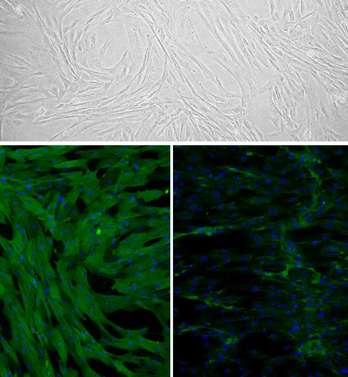
Canine Trabecular Meshwork Cell: CnTMC Top: Phase-contrast, Bottom Left: CnTMC immunostained for alpha-Smooth Muscle Actin (green), Bottom Right: Fibronectin (green), nuclei are stained with DAPI.
Canine Trabecular Meshwork Cells: CnTMC
Canine Trabecular Meshwork Cells (CnTMC) are primary specialized outflow-pathway cells with heterogeneous phenotypes that exhibit unique, hybrid characteristics, derived from canine anterior segment tissue.
Description
Canine Trabecular Meshwork Cells (CnTMC) are primary specialized outflow-pathway cells with heterogeneous phenotypes that exhibit unique, hybrid characteristics, derived from canine anterior segment tissue, specifically cultured in specialized canine trabecular meshwork cell growth medium. Located within a sponge-like connective tissue structure situated near the anterior chamber of the eye, these cells play a critical role in regulating intraocular pressure by controlling the drainage of aqueous humor and modulating outflow resistance. In vivo, trabecular meshwork cells can influence fluid outflow through alterations in contractility and cytoskeletal tension, while also participating in clearance mechanisms, such as the uptake of debris via phagocytic activity. Live cell imaging of the actin cytoskeleton provides valuable insights into cytoskeletal dynamics within CnTMC, supporting ongoing research to identify treatments that target trabecular meshwork contractility, cytoskeletal tension, or extracellular matrix turnover to increase outflow facility and reduce intraocular pressure. Additionally, specific targeting of trabecular meshwork cells has been explored in experimental ophthalmology to help evaluate the delivery efficiency of nanoparticles or gene transfer vectors.
Damage and cellular dysfunction within the trabecular meshwork have significant clinical relevance. For instance, hypoxia can increase DNA methylation and alter gene expression, whereas the normal aging process is often marked by a gradual decrease in cell number and the accumulation of senescent cells. In conditions such as glaucoma — a leading optic neuropathy and a major cause of irreversible blindness characterized by progressive loss of retinal ganglion cells — impaired fluid drainage leads to elevated intraocular pressure. Pathological changes in the trabecular meshwork can include increased fibrosis, fibronectin accumulation, and elevated expression of extracellular matrix cross-linking enzymes, alongside cytoskeletal reorganization and cellular stiffening. Other glaucoma-associated dysfunctions reported in research models involve mitochondrial defects, altered signaling pathways, elevated transforming growth factor-beta 2 (TGF-β2) levels, genomic DNA damage, and oxidative stress.
Understanding trabecular meshwork biology is essential for preserving the functional anatomy of the eye, which relies on transparent structures such as the cornea and lens to properly focus light onto the retina — where specialized photoreceptors, including rods and cones, convert light into neural signals. These signals are subsequently transmitted via the optic nerve to the brain to produce clear vision, emphasizing why maintaining normal outflow facility and preventing pressure-induced damage to the visual pathway remain central goals in vision research.
Canine Trabecular Meshwork Cells (CnTMC) are primary cells derived from canine eyes. CnTMC are cultured in Canine Trabecular Meshwork Cell Growth Medium and are able to propagate to 10 population doublings when cultured under the recommended conditions. Trabecular Meshwork cells are outflow-pathway cells in a sponge-like connective tissue located near the front of the eye and are responsible for regualting eye pressure by controlling drainage of fluid into tubes that flow into the bloodstream. Outflow is mediated by alterations in contractility and tension of TM, which also serve as a self-cleaning filter due to their phagocytic nature. Live cell imaging of the cytoskeleton provides valuable information on actin dynamics in CnTMC. Other research aims to identify treatments that relax TM contraction to increase fluid outflow and lower eye pressure. Specific targeting of TM could also play a clinical role by increasing therapeutic efficacy of nanoparticles for gene delivery. Damage and dysfunction of TM have clinical significance. For instance, hypoxia increases DNA methylation, accompanied by altered gene expression, whereas during the normal aging process, TM number decreases, and senescent cells accumulate.
In Glaucoma, a leading cause of irreversible blindness, decreased fluid outflow causes an elevation of intraocular pressure and progressive loss of retinal ganglion cells. Physical changes to the TM include increased fibrosis, fibronectin accumulation, and expression of ECM cross-linking enzymes. This cytoskeletal reorganization and cell loss causes the TM to become rigid and stiff. Other glaucoma-related dysfunctions include mitochondrial defects, altered signaling pathways, elevated TGF-β2, genomic DNA defects, and oxidation damage.
Details
| Tissue | Normal healthy canine cornea | |
|---|---|---|
| QC | No bacteria, yeast, fungi, mycoplasma, virus | |
| Bioassay | Attach, spread, proliferate in Growth Med | |
| Cryovial | 500,000 CnTMC frozen in Basal Medium w/ 10% FBS, 10% DMSO | |
| Kit | Cryovial frozen CnTMC (Cn634-05), Growth Medium (Cn631-500), Subculture Rgnt Kit (090K) | |
| Proliferating | Shipped in Tsfr Med, flasks or plates | |
| Doublings | At least 12 | |
| Applications | Laboratory research use only (RUO). Not for human, clinical, diagnostic or veterinary use. |
Resources
FAQs
Need More Help?
Visit our comprehensive FAQ page for detailed answers to common questions.
Need More Help?
Visit our comprehensive FAQ page for detailed answers to common questions.
Primary Cell FAQs