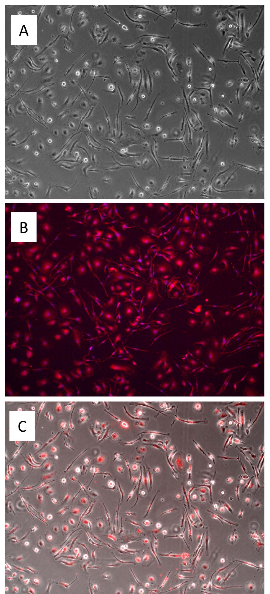
A: MMcg culture on day 4, 100x. B: Immunostaining in MMcg culture on day 4 for F4/80 (red) and DAPI (blue), 200x. C: DiI-Ac-LDL uptake (red) in MMcg culture on day 4, 100x.
Mouse Microglia: MMcg
Mouse Microglia (MMcg) are primary microglial cells typically derived from postnatal mouse brain tissue.
Description
Mouse Microglia (MMcg) are primary microglial cells typically derived from postnatal mouse brain tissue. When revived and plated under optimized cell culture conditions, these primary cells form an adherent culture. Microglia function as the resident macrophage-like immune cells of the central nervous system (CNS). While historically grouped broadly with bone-marrow-derived hematopoietic lineages, current developmental consensus establishes that resident microglia arise primarily from early embryonic yolk-sac myeloid progenitors that seed the developing brain during early development.
In vitro, these primary microglia provide an authentic cell type model for studying neuroimmunology without the drifted phenotypic signatures often observed in immortalized cell lines. Quality control and characterization of these primary microglial cells include verifying the expression of the myeloid/macrophage-associated marker F4/80, alongside functional verification assays evaluating the uptake of fluorescently labeled acetylated low-density lipoprotein (DiI-Ac-LDL). Standard testing is also deployed to ensure lots are free of mycoplasma and other microbial contaminants before research use.
In the living mouse or human CNS, these cells are distributed throughout the parenchyma of the brain and spinal cord. The CNS is dynamically partitioned from systemic circulation by the blood-brain barrier (BBB), leaving resident microglia to function as part of the internal immune surveillance network within the neural tissue.
In a healthy tissue context, homeostatic microglia possess a highly ramified morphology characterized by fine, continuously motile processes. These processes perpetually scan the local microenvironment, working in close contact with surrounding neural components—including neurons, oligodendrocytes, and neighboring astrocyte populations—to monitor tissue health and sustain baseline brain homeostasis.
The primary biological function of microglia is to execute continuous brain immune surveillance, maintain structural homeostasis, and modulate neural circuitry. In a healthy baseline state, these cells manage the targeted clearance of cellular debris resulting from programmed cell death, and they can actively participate in synaptic pruning and contribute to synaptic plasticity.
However, when they encounter microenvironmental disturbances — such as physical brain injury, stroke, or microbial infection — microglia undergo rapid functional shifts, historically described as microglial activation. During this microglial response, the cells can transition from a ramified homeostatic state into an amoeboid, motile cell phenotype. These activated microglia upregulate phagocytic machinery — including pathways that mediate Fc-mediated phagocytosis — and release a complex secretome of cytokines, chemokines, and reactive oxygen species. While a transient response is critical for clearing damaged neural cells, chronic or dysregulated microglial activation is associated with, and can contribute to, accelerated neuroinflammatory tissue damage.
In cellular neurobiology, neuroimmunology, and discovery pharmacology, MMcg cultures serve as a model system to investigate microglial function, analyze neuroinflammatory pathways, and study mechanisms underlying various neurological disorders. Researchers deploy primary microglia to study cellular responses associated with neurodegenerative disease phenotypes. For instance, in modeling Alzheimer’s disease, these cells can be used to evaluate the phagocytic clearance of amyloid-β (Aβ) plaques or to study the transition of homeostatic populations into transcriptionally distinct states, such as disease-associated microglia (DAM) phenotypes, which can be profiled using single-cell RNA sequencing (scRNA-seq) or targeted gene expression panels.
To quantify these cellular behaviors in controlled systems, investigators frequently pair MMcg with a commercial assay kit to measure real-time nitric oxide production, cell proliferation, or chemotactic migration. Primary mouse microglia provide a valuable translational benchmark alongside emerging human microglia models — such as human iPSC-derived microglia or primary human CNS isolates — to decipher potential species-specific differences in receptor expression and signaling networks. By analyzing how candidate immunomodulatory molecules alter the activation kinetics of these primary cells, researchers aim to identify novel checkpoints capable of modulating neuroinflammatory cascades and exploring pathways related to tissue repair across a broad spectrum of neurological diseases.
Mouse Microglia (MMcg) are primary mouse microglia derived from day 1 postnatal CD1 mouse brains. When revived and plated and cultured under recommended conditions, MMcg form adherent culture.
Microglia, originated from the hematopoietic stem cells in bone marrow, are the resident macrophages in the central nervous system (CNS) which is separated from the rest of the body due to the presence of blood-brain barrier. Microglia actively survey the surrounding area and respond by scavenging damaged neural cells, plaques, and infectious agents. Due to their functions in immune response and maintaining homeostasis in the CNS, microglia have been implicated in neurodevelopment, CNS plasticity and repair, neuroinflammation, aging and neurodegeneration, neuropathic pain, and infections.
Characterization: MMcg stain positive for F4/80, and uptake DiI-Ac-LDL
Details
| Tissue | Normal, healthy mouse brain | |
|---|---|---|
| QC | No bacteria, yeast, fungi, mycoplasma | |
| Character | Positive for F4/80 | |
| Bioassay | Plate, spread on CC ware & uptake DiI-Ac-LDL | |
| Cryovial | 1M MMcg frozen in Microglia Cell Freezing Med (043-50) | |
| Kit | Cryovial Frozen MMcg (M8816K-10n), Microglia Cltr Med Kit (M619K-100) | |
| Doublings | N/A | |
| Applications | Laboratory research use only (RUO). Not for human, clinical, diagnostic or veterinary use. |
Resources
FAQs
Need More Help?
Visit our comprehensive FAQ page for detailed answers to common questions.
Need More Help?
Visit our comprehensive FAQ page for detailed answers to common questions.
Primary Cell FAQs