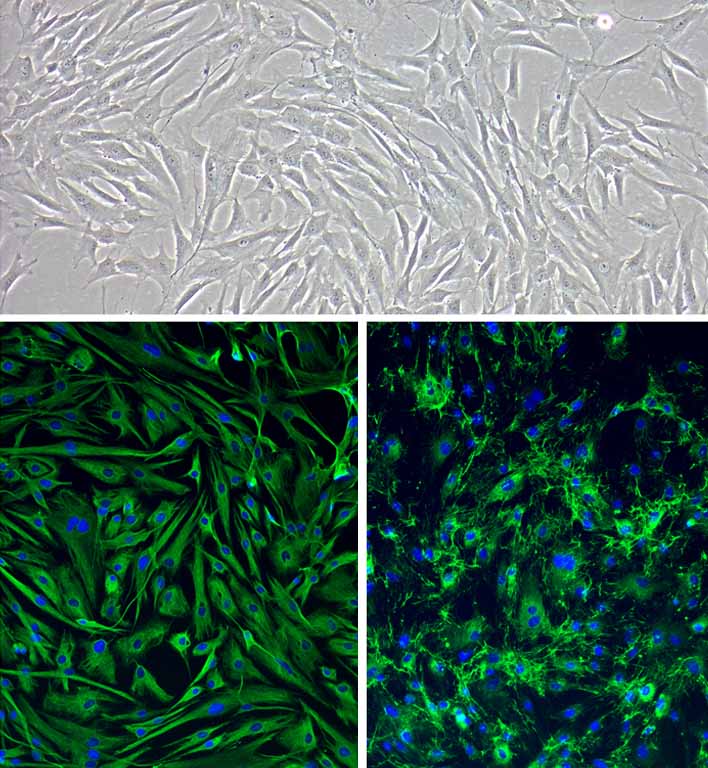
Human Corneal Keratocytes: HCK Top: Phase-contrast, Bottom Left: HCK immunostained for Vimentin (green), Bottom Right: Fibronectin (green), nuclei are stained with DAPI.
Human Corneal Keratocytes: HCK
Human Corneal Keratocytes (HCK) are specialized, neural crest-derived quiescent stromal cell populations that populate the transparent tissue of the eye.
Description
Human Corneal Keratocytes (HCK) — sometimes referred to in the literature as corneal stromal cells or simply human keratocytes — are specialized, neural crest-derived quiescent stromal cell populations that populate the transparent tissue of the eye. While the terms are frequently used interchangeably in broader cell biology contexts, “keratocyte” specifically denotes this quiescent phenotype, whereas “corneal fibroblast” or “myofibroblast” represents distinctly activated, proliferative phenotypes. Unlike an immortalized cell line or transformed cell lines engineered via viral oncogenes (such as hpv16 e6 vectors), primary human keratocytes retain their native phenotypic plasticity, metabolic memory, and precise physiological sensitivity ex vivo. In their resting state, these cells exhibit a distinct dendritic, highly interconnected morphology in vivo. To expand these human primary cells without triggering premature phenotypic drift into active fibroblasts, investigators must utilize an optimized Corneal Keratocyte Growth Medium to maintain baseline gene expression and structural traits before exposing the cells to experimental wounding or differentiation cues.
In the human body, these cells are distributed regularly within the thick, avascular stromal layer of the human cornea. The stroma comprises the vast majority of total corneal thickness and is bordered anteriorly by the stratified corneal epithelium (composed of highly active corneal epithelial cells) and posteriorly by a monolayer of endothelial cells. Within the stroma, HCK are suspended between an intricate arrangement of highly organized, orthogonal collagen lamellae. While early studies focused primarily on surface epithelium or specialized structures like immortalized corneal epithelial/keratinocyte lines, modern ophthalmic research maps the intensive cross-talk that occurs between epithelial cell layers and these deeper stromal cell elements during baseline homeostasis and ocular trauma.
The primary biological function of healthy HCK is to maintain the structural organization, composition, and transparency of the stromal collagen matrix. They synthesize major structural macromolecules—primarily Type I collagen alongside Type V collagen, which acts as a critical regulator of fibril diameter—and produce specialized keratan sulfate proteoglycans such as keratocan, lumican, and decorin that are critical for maintaining uniform fibril spacing and tissue clarity. Furthermore, quiescent HCK express high cytosolic levels of water-soluble crystallins; specifically, robust ALDH3A1 expression helps minimize light scattering and shields deeper ocular structures from ultraviolet radiation.
However, upon injury to the corneal epithelium or mechanical trauma, this homeostasis shifts. Under the influence of epithelial-derived cytokines and localized corneal inflammation, nearby HCK undergo targeted apoptosis or transition into an active repair phenotype. In the classic wound healing cascade, cells follow a sequence from a quiescent keratocyte to a migratory corneal fibroblast, and ultimately into a highly contractile myofibroblast. However, investigators recognize that these cellular transitions can be highly heterogeneous and non-linear, dynamically driven by shifting combinations of biochemical cytokines (such as TGF- and PDGF) and localized biophysical and mechanical cues within the matrix.
In translational ophthalmology and tissue engineering, a primary culture of HCK serves as a critical human-background model for dissecting corneal wound healing, analyzing drug toxicity, and designing biomimetic constructs. Safety pharmacologists deploy HCK to screen topically applied ocular drugs and advanced nanocarriers for localized toxicity, ensuring that novel formulations do not trigger unwanted allergic reactions or cell death. Because the persistent, dysregulated presence of contractile myofibroblasts can lead to the random deposition of disordered matrix components—resulting in corneal fibrosis and a blinding clinical condition known as corneal haze—investigators use HCK to identify small-molecule inhibitors that can halt this fibrotic cascade.
These primary cells are heavily leveraged in regenerative medicine alongside mesenchymal stem cell populations or pluripotent stem cell groups to build tissue-engineered corneal substitutes. By studying how HCK migrate through, adhere to, and organize synthetic polymer hydrogels, bioengineers can optimize 3D-scaffold parameters to repair a damaged stromal matrix and restore complete refractive transparency in vitro.
Details
| Tissue | Normal healthy human cornea | |
|---|---|---|
| QC | No bacteria, yeast, fungi, mycoplasma, virus | |
| Bioassay | Attach, spread, proliferate in Growth Med | |
| Cryovial | 500,000 HCK (1st passage) frozen in Basal Medium w/ 10% FBS, 10% DMSO | |
| Kit | Cryovial frozen HCK (632-05a), Growth Medium (6111-500), Subculture Rgnt Kit (090K) | |
| Proliferating | Shipped in Tsfr Med, psg 1, flasks or plates | |
| Doublings | At least 15 | |
| Applications | Laboratory research use only (RUO). Not for human, clinical, diagnostic or veterinary use. |
Resources
FAQs
Need More Help?
Visit our comprehensive FAQ page for detailed answers to common questions.
Need More Help?
Visit our comprehensive FAQ page for detailed answers to common questions.
Primary Cell FAQs Powered by Bioz