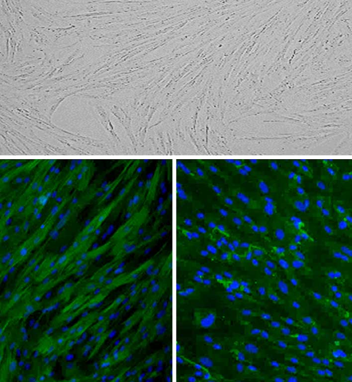
Porcine Trabecular Meshwork Cell: PTMC Top: Phase-contrast, Bottom Left: PTMC immunostained for alpha-Smooth Muscle Actin (green), Bottom Right: Fibronectin (green), nuclei are stained with DAPI.
Porcine Trabecular Meshwork Cells: PTMC
Porcine Trabecular Meshwork Cells (PTMC) are primary cells isolated from pig eyes that are widely used in ophthalmology to model the cellular physiology of the trabecular meshwork, a specialized connective tissue at the anterior segment.
Description
Porcine Trabecular Meshwork Cells (PTMC) are primary cells isolated from pig eyes that are widely used in ophthalmology to model the cellular physiology of the trabecular meshwork, a specialized connective tissue at the anterior segment responsible for maintaining eye health through regulation of aqueous humor outflow. Proper outflow depends on the contractility, cytoskeletal organization, and mechanical properties of trabecular meshwork cells, which in turn influence intraocular fluid dynamics that ultimately affect vision and the health of the retina and optic nerve. Because PTMC capture key aspects of trabecular meshwork cell behavior — such as how cytoskeletal tension, cell–matrix interactions, and extracellular matrix (ECM) homeostasis govern drainage actions — these cells are valuable for studying disease-relevant mechanisms in glaucoma and for evaluating translational strategies aimed at restoring normal fluid outflow.
In glaucoma, decreased fluid outflow contributes to elevated intraocular pressure and progressive vision loss, including damage to the retina and degeneration of the optic nerve. PTMC-based studies support mechanistic investigation of how disease-associated stresses and signaling changes drive structural and functional alterations within the trabecular meshwork. These include increased ECM deposition and fibrotic remodeling, including fibronectin accumulation and changes in enzymes involved in ECM cross-linking, which can shift the tissue toward greater rigidity and reduced drainage capacity. PTMC are also used to explore how cellular stress responses and dysregulated signaling pathways promote trabecular meshwork dysfunction, including changes associated with elevated profibrotic factors such as TGF-β2, mitochondrial defects, altered redox/oxidative balance, and senescence-linked phenotypes that can further impair normal cell actions within the eye.
Beyond characterizing pathology, PTMC enable translational research focused on identifying therapeutic approaches that relax trabecular meshwork contraction or counteract fibrotic remodeling, with the goal of lowering intraocular pressure and preserving retinal and optic nerve function. By linking cell-level mechanisms to functional outcomes in fluid regulation, PTMC provide a practical in vitro platform for testing interventions that could improve therapeutic efficacy in glaucoma-related ophthalmology and contribute to broader translational efforts to protect vision.
Porcine Trabecular Meshwork Cells (PTMC) are primary cells derived from pig eyes. PTMC are cultured in Porcine Trabecular Meshwork Cell Growth Medium and are able to propagate to 10 population doublings when cultured under the recommended conditions. Trabecular Meshwork cells are endothelial-like cells in a sponge-like connective tissue located near the front of the eye and are responsible for regualting eye pressure by controlling drainage of fluid into tubes that flow into the bloodstream. Outflow is mediated by alterations in contractility and tension of TM, which also serve as a self-cleaning filter due to their phagocytic nature. Live cell imaging of the cytoskeleton provides valuable information on actin dynamics in PTMC. Other research aims to identify treatments that relax TM contraction to increase fluid outflow and lower eye pressure. Specific targeting of TM could also play a clinical role by increasing therapeutic efficacy of nanoparticles for gene delivery. Damage and dysfunction of TM have clinical significance. For instance, hypoxia increases DNA methylation, accompanied by altered gene expression, whereas during the normal aging process, TM number decreases, and senescent cells accumulate.
In Glaucoma, a leading cause of irreversible blindness, decreased fluid outflow causes an elevation of intraocular pressure and progressive loss of retinal ganglion cells. Physical changes to the TM include increased fibrosis, fibronectin accumulation, and expression of ECM cross-linking enzymes. This cytoskeletal reorganization and cell loss causes the TM to become rigid and stiff. Other glaucoma-related dysfunctions include mitochondrial defects, altered signaling pathways, elevated TGF-β2, genomic DNA defects, and oxidation damage.
Details
| Tissue | Normal healthy porcine cornea | |
|---|---|---|
| QC | No bacteria, yeast, fungi, mycoplasma, virus | |
| Bioassay | Attach, spread, proliferate in Growth Med | |
| Cryovial | 500,000 PTMC frozen in Basal Medium w/ 10% FBS, 10% DMSO | |
| Kit | Cryovial frozen PTMC (P634-05), Growth Medium (P631-500), Subculture Rgnt Kit (090K) | |
| Proliferating | Shipped in Tsfr Med, flasks or plates | |
| Doublings | At least 17 | |
| Applications | Laboratory research use only (RUO). Not for human, clinical, diagnostic or veterinary use. |
Resources
FAQs
Need More Help?
Visit our comprehensive FAQ page for detailed answers to common questions.
Need More Help?
Visit our comprehensive FAQ page for detailed answers to common questions.
Primary Cell FAQs