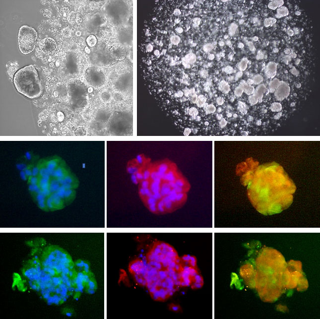
Top Row: 3D culture of AEC2 / i-HAEpC2 organoids embedded in the DISCUSTM system.
Midde Row: Immunostaining of AEC2 / i-HAEpC2 organoids for ACE2/DAPI (green/blue
...Human iPSC-Derived Alveolar Epithelial Cells: AEC2 / i-HAEpC2 (Organoids)
Human iPSC-Derived Alveolar Epithelial Cells: AEC2 / i-HAEpC2 (Organoids) represent a specialized, three-dimensional in vitro model of human Alveolar Type 2 (AT2) epithelial cells.
Description
Human iPSC-Derived Alveolar Epithelial Cells: AEC2 / i-HAEpC2 (Organoids) represent a specialized, three-dimensional in vitro model of human Alveolar Type 2 (AT2) epithelial cells. Produced using a multi-stage directed differentiation protocol, this cell type is engineered to model key stages of human lung development. During differentiation, human induced pluripotent stem cells (iPSCs) are sequentially guided through definitive endoderm (DE cells) and anterior foregut endoderm stages to establish a committed lung progenitor population, which is subsequently transitioned into an alveolar organoid configuration using the DISCUS™ 3D cell culture system.
These human AEC2 models are characterized by distinct gene expression profiles and positive staining for Thyroid Transcription Factor-1 (TTF-1) — a marker for bronchial and alveolar lineages — and Surfactant Protein C (SFTPC), a definitive marker for mature AT2 cells. In this 3D organoid format, the system demonstrates high baseline ACE2 positivity; i-HAEpC2 cells are typically >90% positive for the ACE2 receptor when maintained within the DISCUS™ 3D system. Notably, this high level of ACE2 marker presence is sensitive to the physical microenvironment and is rapidly lost if the cells are subjected to traditional 2D flat-bed plating.
In the living human lung, Alveolar Type 2 (AT2) cells are specialized epithelial components of the distal respiratory zone, nestled within the alveolar epithelium directly adjacent to flat Alveolar Type 1 (AT1) cells. While AT1 cells cover the vast majority of the alveolar surface area to facilitate gas exchange, AT2 cells act as essential caretakers of the alveolus, resting on a basement membrane in close paracrine communication with microvascular endothelial cells and resident interstitial mesenchymal cells.
In response to injury, chronic inflammation, or normal tissue turnover, resident AT2 cells can function as local progenitors. They can exit a quiescent state, re-enter the cell cycle, proliferate, and differentiate into AT1 lung cells to help restore structural barrier integrity. This complex cellular niche and tissue architecture are difficult to replicate in static systems. Complex endodermal tissues, such as the intestine, liver, and distal lung, are widely understood to achieve more representative functional maturation in a 3D structural environment, which serves as the core rationale for utilizing 3D organoid platforms to better reflect the spatial orientation and tissue constraints of native human lung tissue.
The primary biological function of native AT2 cells is to preserve alveolar stability, regulate fluid and ion transport across the epithelium, and participate in localized defense and immune responses. A principal mechanism of these cells is the synthesis, packaging, and secretion of pulmonary surfactant proteins (such as SFTPC), which lower surface tension at the air-liquid interface to prevent alveolar collapse during expiration. Beyond surfactant production, AT2 cells contribute to host defense by releasing a secretome of cytokines and chemokines, which can interact with neighboring immune components, such as resident alveolar macrophage populations, during inflammatory challenges.
When chronic stress or disease-associated pathways disrupt normal AT2 biology, altered cell cycle responses and epithelial distress can contribute to defective tissue repair. In these pathological contexts, epithelial cells can show shifted gene expression patterns and an upregulation of pathways that are consistent with pro-fibrotic signaling. This ongoing distress can cross-talk with neighboring mesenchymal cells, which can contribute to the progressive matrix deposition and tissue remodeling observed in progressive pulmonary fibrosis models.
As a Research Use Only (RUO) platform, i-HAEpC2 organoids provide a human-background model system to investigate distal lung biology, organoid formation, and the mechanisms of chronic lung disease without requiring scarce primary human AEC2 or lung tissue samples. Researchers deploy these 3D systems to evaluate changes in gene expression and model localized epithelial responses during injury. Because these organoids natively maintain high ACE2 positivity when cultured in the proper 3D format, they are useful for investigating respiratory pathogen interactions and studying ACE2-dependent cell entry-related biology.
Furthermore, these iPSC-derived alveolar models can be used in research workflows to examine the molecular mechanics of progressive lung fibrosis, including idiopathic pulmonary fibrosis (IPF). Investigators utilize the platform to analyze how specific environmental stimuli or pro-inflammatory cytokines alter cell cycle kinetics, evaluate targeted gene sets, and study anti-fibrotic interventions. Finally, these human-background cell lots serve as a scalable benchmark in experimental designs requiring cross-species comparisons, allowing researchers to evaluate human-specific epithelial responses alongside established mouse lung baselines.
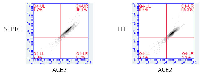 Endodermal tissues such as intestine, liver and lung reach full function only in a 3-D environment. Cell Applications’ i-HAEpC2 are typically >90% positive for ACE2 receptor. The marker is only present when cells are in the DISCUSTM 3D system and is rapidly lost if subjected to 2D plating.
Details
| Source | Derived from Human iPSC of a single donor | |
|---|---|---|
| QC | No bacteria, yeast, fungi, mycoplasma, virus | |
| Bioassay | Positive for ACE2 staining. | |
| Cryovial | 500,000 AEC2 / i-HAEpC2 (1st passage) in freezing medium | |
| Kit | Cryovial frozen AEC2 / i-HAEpC2 (i538-05), Gr Med Kit (i51103K-100), i-HAEpC2 DISCUSTM Gel Kit (DISC1054K), Wash Buffer (077-25), DISCUSTM Dissociation Solution (DISC1050), Non-Stick Solution (1025-05). | |
| Cultured | Contact Cell Applications tech support for custom cultures | |
| Doublings | The cells cannot be passaged | |
| Applications | Laboratory research use only (RUO). Not for human, clinical, diagnostic or veterinary use. |
Resources
FAQs
Need More Help?
Visit our comprehensive FAQ page for detailed answers to common questions.
Need More Help?
Visit our comprehensive FAQ page for detailed answers to common questions.
Primary Cell FAQs Powered by Bioz