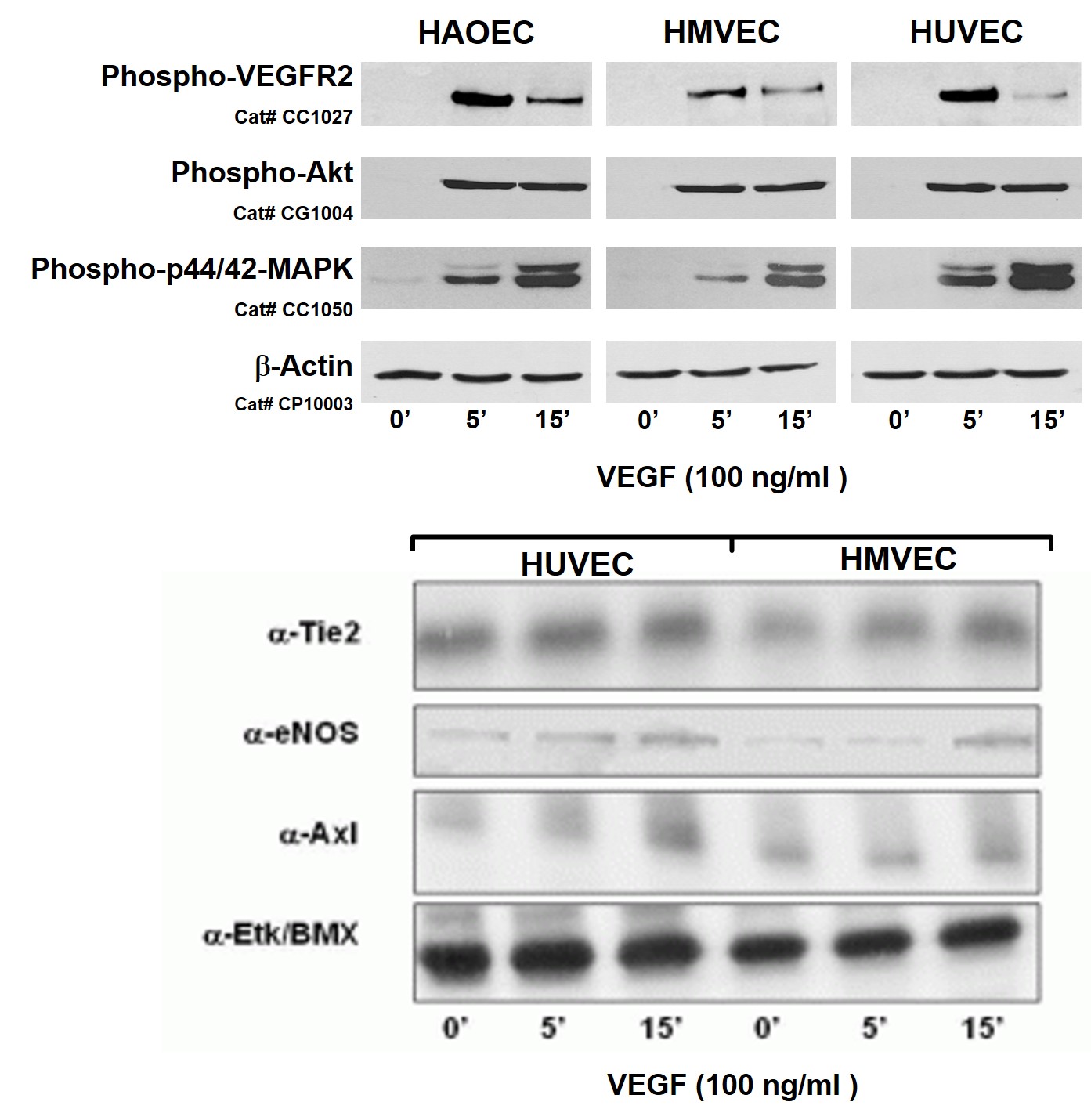
Pre-Screened Endothelial Cells Top: Activation of VEGFR-2 signaling in HAOEC, HUVEC and HMVEC by 100 ng/ml VEGF for 0, 5 or 15 minutes. VEGFR-2 autophosphorylation probed w/ phospho-VEGFR-2 (Y1175) specific Ab, Akt activation detected by phospho-Akt (S473) specific antibody, and MAP kinase activation detected using phospho-p44/42 MAP kinase (T202/Y204) Ab. Bottom: Pre-screened
...Human Aortic Endothelial Cells: S-HAOEC: Pre-Screened
Human Aortic Endothelial Cells: Pre-Screened (S-HAOEC) — commercially validated lots of a primary human aortic endothelial cell line — are isolated from the tunica intima of the human aorta.
Description
Human Aortic Endothelial Cells: Pre-Screened (S-HAOEC) — commercially validated lots of a primary human aortic endothelial cell line — are isolated from the tunica intima of the human aorta. As the largest elastic artery in the cardiovascular system, the aorta is continuously subjected to high-pressure pulsatile blood flow, making these vascular endothelial cells highly specialized to handle significant cyclic strain. To mitigate the pronounced donor-to-donor variability and drifting signaling kinetics typical of un-validated primary tissue isolates, S-HAOEC lots undergo strict quality control. They are pre-screened to verify that they maintain specific characteristics, regular morphology, and are free of contaminants like mycoplasma. Cultured in optimized endothelial cell growth media with an advanced formulation, these primary endothelial cells provide a translationally rigorous model for investigating atherosclerosis, diabetes, and systemic vascular disease.
The pre-screening protocol evaluates the cellular machinery of S-HAOEC to ensure the macrovascular-specific functions (such as high-pressure mechanical responses and robust VEGFR-2 cascades) remain intact, distinct from venous networks or microvascular niches:
- VEGFR-2 Activation: VEGFR-2 activation by VEGF is a well-established pathway for endothelial cell survival and proliferation. Upon ligand binding, vascular endothelial growth factor induces receptor homodimerization and autophosphorylation of VEGFR-2, propagating downstream signals through the Akt and p44/42 MAPK (ERK1/2) pathways.
- Endothelial Markers: Validated lots maintain the baseline gene expression of definitive markers and co-regulators, including von Willebrand factor (vWF), which stabilizes Factor VIII and mediates platelet adhesion. Compared to vascular smooth muscle cells or general human dermal fibroblasts, S-HAOEC preserve a native arterial endothelial profile that is heavily documented across peer-reviewed scientific references and industrial white papers.
Because these cells line a high-pressure macrovascular environment, they are uniquely suited for modeling disturbed or altered shear-induced pathology and endothelial dysfunction (or endothelium dysfunction).
- Mechanotransduction: Laminar shear stress (~10-20 dyn/cm2) aligns endothelial cells in the direction of flow and upregulate protective nitric oxide signaling. Ando J, et al. demonstrated that laminar shear stress upregulates KLF2 and eNOS in endothelial cells, which provides an essential control when contrasting normal physiology against the turbulent, oscillatory shear stress patterns (~ 0.5-5 dyn/cm2) at aortic bifurcations that trigger atherosclerosis.
- Disease Mechanisms: These cell models are highly effective for simulating the impact of hypertension, where high shear induces oxidative stress, and diabetes, where hyperglycemia and advanced glycation end-products (AGEs) impair baseline functions. Under these chronic stresses, cells upregulate cellular adhesion molecules (such as VCAM-1, ICAM-1, and E-selectin), facilitating leukocyte docking and modeling the focal inflammation that drives plaque formation in the aorta and coronary artery (and wider human coronary artery) networks, contributing to coronary artery disease.
The robust handling and uniform differentiation profiles of S-HAOEC make them a foundational workhorse across diverse primary cells platforms for biotechnology applications.
- Vascular Regulation: Beyond basic barrier properties, S-HAOEC actively regulate vascular tone via the balanced release of nitric oxide, prostacyclin (PGI2), and endothelin-1 (ET-1), while maintaining a thromboresistant surface through the expression of thrombomodulin and tissue factor pathway inhibitor (TFPI). They also coordinate sprouting angiogenesis via coordinated VEGF and Notch signaling.
- Engineered Tissues: Bioengineers leverage S-HAOEC to assemble complex 3d endothelialized engineered tissues and vascularize custom engineered tissues. These platforms allow researchers to evaluate the hemocompatibility of next-generation vascular implants and build high-throughput screening pipelines to identify small molecules and evaluate novel disease treatments for complex cardiovascular disease states.
Human Aortic Endothelial Cells: Pre-Screened for Angiogenesis & VEGF Signaling – The most commonly used human endothelial cells are Human Umbilical Vein Endothelial Cells, (HUVEC), Human Microvascular Endothelial Cells (HMVEC) and Human Aortic Endothelial Cells (HAOEC). At Cell Applications, select lots of HUVEC, HMVEC, HLMVEC, and HAOEC have been pre-screened to demonstrate stimulation-dependent angiogenesis and key endothelial cell signaling pathways (phosphorylation of VEGFR, Akt, MAPK, and expression of Tie2, eNOS, Axl and Etk/Bmx. Other cell types can be pre-screened by customer request.
VEGF-Stimulated Signaling in Pre-Screened Endothelial Cells -VEGF receptor-2 (VEGFR-2) is a major VEGF of endothelial cells. VEGFR-2-mediated signaling plays a critical role in angiogenesis, including regulation of proliferation, differentiation, cell movement, and survival of endothelial cells. VEGF-induced receptor dimerization triggers activation of VEGFR-2 tyrosine kinase and autophosphorylation at a specific set of tyrosine residues, which serve as docking sites for downstream signaling components leading to activation of downstream signaling molecules, including phosphorylation of Akt and p44/42-MAPK.
Details
Expression of Signaling Biomarkers in Pre-Screened Endothelial Cells. Prescreened Endothelial Cells from Cell Applications, Inc. express the following important biomarkers:
- Tie2, a receptor tyrosine kinase critical for the angiogenic remodeling, sprout formation, survival of endothelial cells and vessel stabilization processes
- eNOS (endothelial Nitric Oxide Synthase), the enzyme that producse NO which is an important signaling molecule that regulates a diverse range of physiological events and is required for normal endothelial function
- Axl, a receptor tyrosine kinase whose ligand is the survival factor Gas6 (growth arrest-specific gene 6 product), is implicated in cell survival, leukocyte transmigration and neointima formation
- Etk/Bmx (Endothelial/epithelial Tyrosine Kinase), a member of the Btk family, participates in signal transduction stimulated by growth factor receptors, cytokine receptors, G-protein-coupled receptors, antigen receptors, and integrins, and has been implicated in cell adhesion, migration, proliferation and survival.
Resources
FAQs
Need More Help?
Visit our comprehensive FAQ page for detailed answers to common questions.
Need More Help?
Visit our comprehensive FAQ page for detailed answers to common questions.
Primary Cell FAQs Powered by Bioz