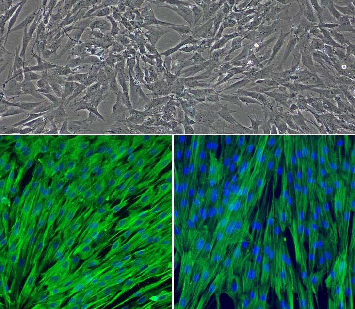
Top: Porcine Cardiac Valve Interstitial Cells (PCVIC). Bottom left: PCVIC stained for vimentin (in green) and DAPI (in blue). Bottom right: PCVIC stained for α-smooth muscle actin (in green) and DAPI (in blue).
Porcine Cardiac Valve Interstitial Cells: PCVIC
Porcine Cardiac Valve Interstitial Cells (PCVIC) are the heterogeneous mesenchymal cell type residing within the core layers of cardiac valves.
Description
Porcine Cardiac Valve Interstitial Cells (PCVIC) — commonly referred to in heart valve biology as valvular interstitial cells (VIC) — are the predominant, heterogeneous mesenchymal cell type residing within the core layers of cardiac valves. As a true primary cell population rather than a modified cell line, these connective tissue components maintain the functional characteristics of their tissue of origin, making them a useful translational model. In a resting physiological state in vitro, primary PCVIC typically display a spindle-shaped morphology and exhibit distinct immunocytochemical characterization patterns, staining positive for vimentin, while the expression of α-smooth muscle actin (α-SMA) is low or absent in many quiescent VICs and serves as a primary marker for myofibroblastic activation.
To preserve their native quiescent cell phenotype and avoid spontaneous activation in vitro, these primary cells require highly optimized, species-specific culture conditions. They are typically maintained in a specialized Porcine Cardiac Valve Interstitial Cell Growth Medium according to protocols. Because their baseline signaling networks and functional profiles share many features with human heart valves, these primary porcine cells serve as an excellent in vitro model system, minimizing the need to rely on rare human valve tissues. They can also be utilized as a comparative benchmark alongside mesenchymal derivatives of pluripotent stem cell lines, including induced pluripotent stem cell (iPSC) and embryonic stem cell models.
In the living porcine or human heart, these interstitial cells are embedded throughout the complex extracellular matrix of the aortic valve leaflets (or aortic valve cusps). The aortic valve tissue is composed of three distinct structural layers—the fibrosa, spongiosa, and ventricularis—which are carefully organized to withstand intense, repetitive hemodynamic stresses during the cardiac cycle. Within these layers, PCVIC reside in close proximity to a specialized surface monolayer of valvular endothelial cells.
While aortic valve endothelial cells directly interface with blood flow to regulate paracrine signaling and manage permeability, the underlying interstitial cell population manages the structural maintenance and remodeling of the deeper matrix layers. While these cardiac cells share a mesenchymal lineage with other stromal populations throughout the body, they possess unique mechanosensitive properties adapted specifically to withstand intense pulsatile pressures within the mammalian heart.
The primary biological function of healthy PCVIC is to maintain the architectural integrity, mechanical compliance, and continuous turnover of the valvular extracellular matrix. Under normal physiological conditions, these cells regulate the synthesis and spatial organization of essential structural elements, such as collagen, elastin, and glycosaminoglycans, which allow the heart valve to flex and seal millions of times over a lifespan.
However, when chronic mechanical strain, pro-inflammatory cytokines, or disturbed shear stress disrupt this homeostatic balance, PCVIC can undergo a pathobiological shift known as myofibroblastic activation. In this activated state, the cells upregulate α-smooth muscle actin (α-SMA) expression, increase cellular contractility, and alter their matrix-remodeling enzymes. In progressive disease models, this persistent activation can contribute to osteogenic differentiation and calcification, leading to micro-calcification, severe tissue remodeling, and compliance loss. This cellular pathobiology is consistent with the initiation of valve calcification, a foundational phase in calcific aortic valve disease (CAVD), which can ultimately culminate in clinical aortic valve stenosis (aortic stenosis).
In cardiovascular research, cell biology, and discovery pharmacology, PCVIC serve as a large-animal platform to investigate the molecular mechanisms driving aortic valve disease and to explore novel therapeutic interventions. Researchers frequently deploy PCVIC in 3D cell culture systems—such as encapsulating them within a functional collagen gel matrix—to study cell-matrix interactions, monitor real-time contraction kinetics, and track changes in gene expression under controlled mechanical loads. The platform is utilized in cardiovascular studies, including those published in journals such as Circulation Research, to evaluate how specific biochemical stimuli alter calcification pathways.
PCVIC provide a crucial translational testbed for evaluating new strategies to treat patients suffering from advanced aortic valve calcification. Because there are currently limited pharmaceutical options to reverse severe leaflet calcification or stenosis, advanced calcification typically leaves patients dependent on high-risk surgical aortic valve replacement or transcatheter valve replacement procedures. Investigators leverage these primary cell lots to examine pathways involved in osteogenic differentiation, evaluate targeted small-molecule or biologic interventions, and study tissue-engineered biomaterials designed to improve the long-term durability of next-generation bioprosthetic human valve substitutes.
Porcine Cardiac Valve Interstitial Cells (PCVIC) are a heterogeneous population of specific cell types responsible for maintaining the structural integrity and normal functioning of the valve. Primary culture of valve interstitial cells is useful in exploring cellular and molecular pathogenesis of heart valve disease and discovering new therapeutic targets for cardiovascular diseases.
PCVIC from Cell Applications, Inc. are derived from porcine heart and cultured in Porcine Cardiac Valve Interstitial Cell Growth Medium. PCVIC can propagate to 10 population doublings when cultured under the recommended conditions and provide an excellent model system to study heart valve function and pathobiology.
Characterization: Positive for α-smooth muscle actin and vimentin by ICC staining.
Details
| Tissue | Normal healthy porcine heart | |
|---|---|---|
| QC | No bacteria, yeast, fungi, mycoplasma | |
| Character | Positive for α-smooth muscle actin and vimentin | |
| Bioassay | Attach, spread, proliferate in Growth Med | |
| Cryovial | 500,000 PCVIC (1st passage) frozen in Basal Medium w/ 10% FBS, 10% DMSO | |
| Kit | Cryovial frozen PCVIC (P370-05n), Growth Medium (P3113-500), Subculture Rgnt Kit (090K) | |
| Proliferating | Shipped in Gr Med, 2nd psg (flasks or plates) | |
| Doublings | At least 15 | |
| Applications | Laboratory research use only (RUO). Not for human, clinical, diagnostic or veterinary use. |
Resources
FAQs
Need More Help?
Visit our comprehensive FAQ page for detailed answers to common questions.
Need More Help?
Visit our comprehensive FAQ page for detailed answers to common questions.
Primary Cell FAQs