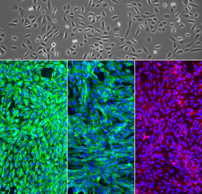
Human Renal Glomerular Epithelial Cells: HRGEpC
Human Renal Glomerular Epithelial Cells (HRGEpC) are highly specialized, largely post-mitotic in vivo visceral epithelial cell types that envelop the glomerular capillary tuft.
Description
Human Renal Glomerular Epithelial Cells (HRGEpC), also commonly referred to as human podocytes, are highly specialized, largely post-mitotic in vivo visceral epithelial cell types that envelop the glomerular capillary tuft. Positioned within Bowman’s capsule, these cells are essential components of the glomerular filtration barrier (GFB). This barrier functions as an integrated unit, where size and charge selectivity are governed by the synergy of the fenestrated endothelial cell layer, the glomerular basement membrane (GBM), and the foot processes of the HRGEpC.
While HRGEpC are distinct, understanding their pathology requires contextual contrast with other defined populations, such as proximal tubule epithelial cells, distal tubule/collecting duct epithelial cells, and other human renal epithelial cells. Comparing these renal epithelial cells—including renal cortical epithelial cells—is essential to mapping the specific functional segment responses that define global kidney health.
In vivo, HRGEpC extend interdigitating foot processes that form the slit diaphragm, a specialized junction integral to the filtration process. Because immortalized lines often exhibit unstable cytoskeletal dynamics, primary HRGEpC are often preferred for high-fidelity studies. These models are vital for mapping the progression of renal disease, including the mechanical and metabolic pathways involved in diabetic nephropathy, where the structural integrity of the GFB is compromised.
Effective research relies on delineating podocyte-specific responses from those of tubular epithelial cells located in the proximal tubule. Differentiating these cell types is critical for the following applications:
-
Mechanosensing and Shear Stress: HRGEpC function as mechanosensors in the high-pressure capillary network, dynamically adjusting their actin cytoskeleton to cyclic stretch. Investigators study these cells to understand how mechanical strain modulates slit diaphragm stability, contrasting these dynamics with the physiological stressors experienced by the renal proximal tubule during filtrate reabsorption.
-
Nephrotoxicity and Injury Mechanisms: The study of nephrotoxicity is highly context-dependent. While many studies focus on tubular epithelial cells in the proximal tubule due to their active role in drug uptake and metabolism, HRGEpC are specific targets for agents that disrupt the GFB. Researchers compare the responses of HRGEpC to those of cells in the renal proximal tubule or the cortical collecting duct to determine the precise localization and mechanism of toxic injury.
-
Advanced Microfluidic Modeling: To recapitulate the kidney’s architecture, microfluidic Glomerulus-on-a-Chip platforms seed HRGEpC on one side of a porous membrane, with renal glomerular endothelial cells on the opposite channel. This allows for the investigation of hemodynamic forces, facilitating the study of how glomerular health interacts with other segments, including the proximal tubule and the cortical collecting duct.
By utilizing primary HRGEpC models, investigators can better dissect the molecular drivers of renal disease and diabetic nephropathy, ensuring that interventions are evaluated within a physiologically accurate framework that accounts for the complex, integrated nature of the filtration barrier.
Details
| Tissue | Normal healthy human kidney | |
|---|---|---|
| QC | No bacteria, yeast, fungi, mycoplasma, virus | |
| Character | Nephrin, pan-CK, Vimentin positive | |
| Bioassay | Attach, spread, proliferate in Growth Med | |
| Cryovial | 500,000 HRGEpC (2nd passage) frozen in Basal Med w/ 10% FBS, 10% DMSO | |
| Kit | Cryovial frozen HRGEpC (942-05n), Gr Med (911-500), Subculture Rgnt Kit (090K) | |
| Proliferating | Shipped in Gr Med, 3rd psg (flasks or plates) | |
| Doublings | 2-3 | |
| Applications | Laboratory research use only (RUO). Not for human, clinical, diagnostic or veterinary use. |
Resources
FAQs
Need More Help?
Visit our comprehensive FAQ page for detailed answers to common questions.
Need More Help?
Visit our comprehensive FAQ page for detailed answers to common questions.
Primary Cell FAQs Powered by Bioz