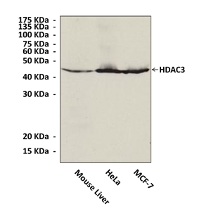
Anti-HDAC3: HDAC3 Mouse Monoclonal Antibody
HDAC3 Mouse Monoclonal Antibody: HDAC3 Mouse Monoclonal Antibody
Size: 100 ul
Price: $302.00
Description
Vertebrates express at least 18 distinct HDACs which have been grouped into four classes based on their similarity with yeast HDACs.2 Class I HDACs (HDACs 1, 2, 3 and 8) are composed primarily of a catalytic domain. These HDACs are ubiquitously expressed, localized to the nucleus, and serve as transcriptional repressors. Class II HDACs (HDAC 4, 5, 6, 7, 9 and 10) are larger proteins with an extended N-terminus extension involved in protein-protein interactions. These HDACs are further sub-classified into Class IIa (HDACs 4, 5, 7, and 9) and IIb (HDACs 6 and 10). In contrast to Class I HDACs, Class II HDACs display stage and tissue-specific expression. These HDACs are able to shuttle in and out of the nucleus in response to certain cellular signals.
Phosphorylation of these HDACs at two N-terminal serine residues by the calcium-calmodulin-dependent kinase (CaMK) creates a docking site for 14-3-3 proteins, resulting in their export out of the nucleus. HDAC11 is the sole member of the Class III HDAC family. The fourth class of deacetylases, called sirtuins (SIRT1-7), has catalytic domains similar to the yeast NAD+-dependent deacetylase Sir2. While serving as HDACs in yeast, in mammalian cells SIRTs are involved in the deacetylation of proteins other than histones, and are therefore not considered to be “classical HDACs.”
HDAC3 is located in both nuclear and cytoplasmic compartments as well as at the plasma membrane. It is required for cell growth and involved in the apoptotic process of almost all cell types via the regulation of pro-apoptotic genes. Moreover, HDAC3 has been suggested to have a role in the cytoplasm, notably in signal transduction since it is a substrate of the membrane associated tyrosine kinase Src. This enzyme plays a critical role in development, inflammation and metabolism. It is present in specific complexes containing members of the nuclear receptor co-repressor family N-CoR/SMRT. HDAC3 is responsible for the deacetylation of lysine residues on the N-terminal part of the core histones that correlates with epigenetic repression. It can also target non-histone proteins. The activity of HDAC3 is regulated by the phosphorylation of the Ser424 residue of the protein and CK2 and PP4 are responsible for this regulation.3 The cleavage of the HDAC3 protein is another type of regulation affecting this enzyme.4
2. Yang X and Seto E: Oncogene 26:5310–5318, 2007
3. Zhang, X. et al: Gene Dev. 19:827-39, 2005
4. Escaffit, F. et al: Mol. Cell. Biol. 27:554-67, 2006
Details
| Cat.No.: | CC10027 |
| Antigen: | Raised against a short peptide from human HDAC3 sequence. |
| Isotype: | Mouse IgG1 |
| Species & predicted species cross- reactivity ( ): | Human, Mouse, Rat |
| Applications & Suggested starting dilutions:* | WB 1:1000 IP n/d IHC n/d ICC n/d FACS n/d |
| Predicted Molecular Weight of protein: | 49 kDa |
| Specificity/Sensitivity: | Detects endogenous HDAC3 proteins without cross-reactivity with other family members. |
| Storage: | Store at -20°C, 4°C for frequent use. Avoid repeated freeze-thaw cycles. |
*Optimal working dilutions must be determined by end user.
