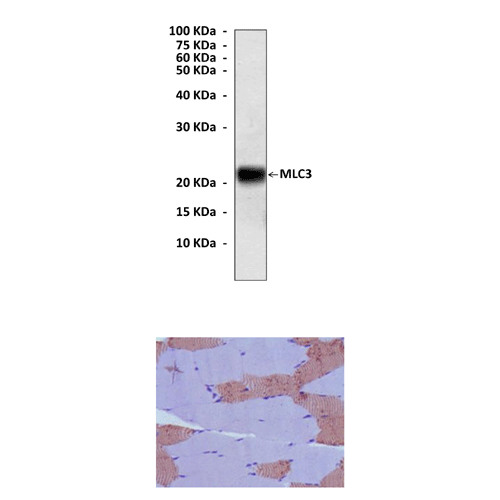
Anti-MLC3: Mouse Myosin Light Chain 3 Antibody
Mouse Myosin Light Chain 3 Antibody: Mouse Myosin Light Chain 3 Antibody
Size: 100 ul
Price: $383.00
Description
Myosin movement can be regulated by phosphorylation of the regulatory light chain of myosin (RLC). This RLC is phosphorylated by Ca2+/calmodulin-dependent myosin light chain kinase (MLCK) or PKC at Ser19 of RLC, which resulted in increased actin-stimulated myosin MgATPase activity. The phosphorylation also increased Ca2+-stimulated myofibrillar MgATPase activity upon substitution of the phosphorylated myosin into myofibrils. This will enable the myosin crossbridge to bind to the actin filament and allow contraction to begin (through the crossbridge cycle). Thus, phosphorylation of RLC by Ca2+/calmodulin-dependent MLCK is a critical step in the initiation of smooth muscle and non-muscle cell contraction.2 In addition, Myosin regulatory light chain (RLC) phosphorylation has been implicated in Rho-mediated stress fiber formation. It was reported that gamma-PAK, which is activated by the GTP-binding proteins Cdc42 and Rac, catalyses phosphorylation of intact non-muscle myosin II and isolated recombinant RLC. Phosphorylation is Ca2+/calmodulin-independent and Ser-19 is the only phosphorylation site modified by gamma-PAK. Similar to MLCK, Arg-16 is required for interaction of gamma-PAK with the substrate. It was suggested that myosin II activation by the p21-activated family of kinases may be physiologically important in regulating cytoskeletal organization.3
In addition, MLC2 participates in various cell signaling regulations in non-muscle cells. It is known that cells exert force propelling the cell forward by contraction of the actin cytoskeleton through activation of myosin II. The actin-myosin II interaction in non-muscle cells is regulated by the phosphorylation of MLC2 at serine-19 too. MLC2 dephosphorylation can induce apoptosis and inhibitor of MLCK can abrogate MLC2 phosphorylation, cell polarization and migration. MLC2 is also involved in the activation of mid-G1 phase cyclin D1 expression. It has been reported that hyperphosphorylated MLC2 induces stress fiber formation and integrin clustering that link cell surface cytoskeletal proteins such as FAK to actin and activates FAK downstream signaling.4
2. Noland, T. A. & Kuo, J.F.: Biochem. Biophy. Res. Commun. 193:254-60, 1993
3. Yoneda, A. et al: J. Cell Biol. 170:443-53, 2005
4. Rek, K et al: J. Biol. Chem. 283:35598-605, 2008
Details
| Cat.No.: | CP10177 |
| Antigen: | Purified recombinant human MLC3 fragments expressed in E. coli. |
| Isotype: | Mouse IgG1 |
| Species & predicted species cross- reactivity ( ): | Human, Mouse, Rat |
| Applications & Suggested starting dilutions:* | WB 1:1000 IP n/d IHC 1:100 ICC n/d FACS n/d |
| Predicted Molecular Weight of protein: | 22 kDa |
| Specificity/Sensitivity: | Detects endogenous MLC3 proteins without cross-reactivity with other family members. |
| Storage: | Store at -20°C, 4°C for frequent use. Avoid repeated freeze-thaw cycles. |
*Optimal working dilutions must be determined by end user.
