Anti-p44/Erk1: Mouse p44/Erk1 Antibody
Mouse p44/Erk1 Antibody: Mouse p44/Erk1 Antibody
Size: 100 ul
Price: $457.00
Description
ERK1 and ERK2 are proteins of 44 and 42 kDa that are nearly 85% identical overall, with much greater identity in the core regions involved in binding substrates. ERK1 and ERK2 are activated by a pair of closely related MEKs, MEK1 and MEK2. The two phosphoacceptor sites, tyrosine and threonine, which are phosphorylated to activate the kinases, are separated by a glutamate residue in both ERK1 and ERK2 to give the motif TEY(Thr202/Tyr204 and Thr185/Tyr187 for p44 and p42 respective) in the activation loop. Once activated, Erk1/2 rapidly translocated into the nucleus, where Erk1/2 activates downstream signaling components including Elk-1, although activated Erk1/2 also phosphorylated numerous substrates on (S/T)P sites in all cellular compartments.2 The Erk1/2 was inactivated by dephosphorylation through action of MAPK phosphotase 1 and 2 in nucleus.3
2. Farooq A & Zhou MM: Cell. Signal. 16:769-779, 2004.
3. Pouyssegur J et al.: Biochem. Pharm. 64:755-763, 2002.
Details
| Cat.No.: | CP10409 |
| Antigen: | Raised against recombinant human p44/Erk1 fragments expressed in E. coli. |
| Isotype: | Mouse IgG1 |
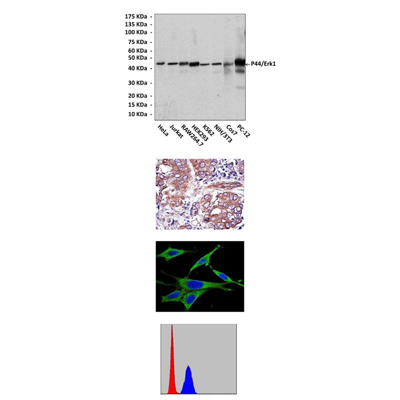
| Species & predicted species cross- reactivity ( ): | Human, Mouse, Rat |
| Applications & Suggested starting dilutions:* | WB 1:1000 IP 1:50 IHC 1:50 – 1:200 ICC 1:50 – 1:200 FACS 1:50 – 1:200 |
| Predicted Molecular Weight of protein: | 44 kDa |
| Specificity/Sensitivity: | Detects p44/Erk1 proteins in various cell lysate. |
| Storage: | Store at -20°C, 4°C for frequent use. Avoid repeated freeze-thaw cycles. |
*Optimal working dilutions must be determined by end user.