Anti-Smad6: Mouse Smad6 Antibody
Mouse Smad6 Antibody: Mouse Smad6 Antibody
Size: 100 ul
Price: $413.00
Description
Smad proteins transduce signals from transforming growth factor-beta (TGF-beta) superfamily ligands. It has been demonstrated that accessory/scaffolding proteins interact with the type I and II receptors and/or the Smads. One example is SARA (Smad anchor for receptor activation), a cytoplasmic protein that specifically interacts with non-activated Smad2 and the receptor complex, thus forming a bridge between the receptor and Smad2 and assisting in the specific phosphorylation of Smad2 by the type I receptor. The mechanism that organises such Smad signalling centres and its links to receptor endocytosis, degradation and signalling crosstalk could provide cell-context specificity, allowing differential regulation of the basic Smad pathway.2
Phosphorylation of the C-terminal serine residues in R-Smads by type I receptor kinases is a crucial step in TGF-beta family signaling. The two most C-terminal serine residues become phosphorylated and, together with a third, non-phosphorylated serine residue, form an evolutionarily conserved SSXS motif in all R-Smads. TGF-beta and activin receptors phosphorylate Smad2 and Smad3, and BMP receptors phosphorylate Smad1, Smad5 and Smad8. Other kinases might also phosphorylate the Smads, which include MAPK, CaMK II and PKC.3 The phosphorylation at Ser423 and Ser425 of Smad3, which triggers dissociation of Smad3 from its receptors to form a complex with Smad4 and accumulate in the nucleus. Unphosphorylated Smad proteins exist primarily as monomers, and upon phosphorylation, R-Smads form homo-oligomers, which quickly convert to hetero-oligomers containing the Co-Smad, Smad4 and are imported to the nucleus. Nuclear Smad oligomers bind to DNA and associate with transcription factors to regulate expression of target genes. Alternatively, nuclear R-Smads associate with ubiquitin ligases and promote degradation of transcriptional repressors, thus facilitating target gene regulation by TGF-beta. Smads themselves can also become ubiquitinated and are degraded by proteasomes. Finally, the inhibitory Smads (I-Smads) block phosphorylation of R-Smads by the receptors and promote ubiquitination and degradation of receptor complexes, thus inhibiting signaling.4
2. Dijke, P.T. & Hill, C.S. : Trends Biochem. Sci. 29:265-73, 2004
3. Massagué, J. et al: Gene Dev. 19:2783-10, 2005
4. Miyazawa, K. et al: Gene. Cell.7:1191-1204, 2002
Details
| Cat.No.: | CP10258 |
| Antigen: | Purified recombinant human Smad6 fragments expressed in E. coli. |
| Isotype: | Mouse IgG1 |
| Species & predicted species cross- reactivity ( ): | Human, Mouse, Rat |
| Applications & Suggested starting dilutions:* | WB 1:1000 IP 1:50 IHC n/d ICC n/d FACS n/d |
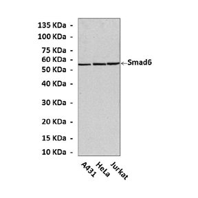
| Predicted Molecular Weight of protein: | 54 kDa |
| Specificity/Sensitivity: | Detects endogenous Smad6 proteins without cross-reactivity with other related proteins. |
| Storage: | Store at -20°C, 4°C for frequent use. Avoid repeated freeze-thaw cycles. |
*Optimal working dilutions must be determined by end user.