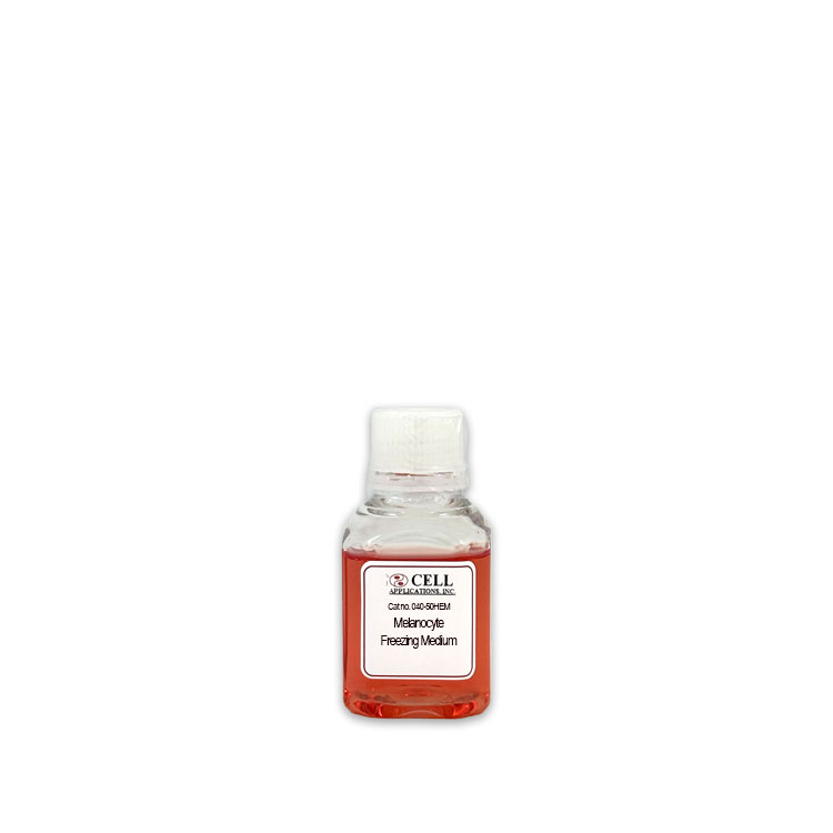
Human Melanocyte Freezing Medium
Freezing Medium for HEM: Freezing Medium optimized for the unique needs of Human Epidermal Melanocytes (HEM).
Size: 50 ml
Price: $79.00
Description
Human Melanocyte Freezing Medium is a specialized formulation designed to help preserve the viability of primary human cells during freeze storage. By supporting cellular integrity and helping reduce cryoinjury, this medium helps maintain delicate melanocyte cultures for future use after thawing.
In the human body, melanocytes are specialized, dendritic pigment-producing cells derived from the neural crest, a neuroectodermal lineage. These cells are found primarily in the basal layer of the epidermis, where they interact with neighboring keratinocytes. Their main function is to synthesize melanin within specialized organelles called melanosomes, and this pigment is transferred to surrounding keratinocytes to help protect skin from ultraviolet radiation.
Studying melanogenesis, pigmentation disorders, photoaging, and melanoma in vitro requires access to healthy primary cultures. Because primary human cells can be sensitive to cryopreservation, Human Melanocyte Freezing Medium helps create a protective environment for freezing and storage in liquid nitrogen for later experimental use.
Freezing Media (also referred to as cryopreservation media) is a specialized category of cell culture formulation engineered to protect cells from ice crystal formation and osmotic shock during the freezing and thawing processes.
The primary biological and physical function of freezing media is to manage the thermodynamic and osmotic transitions that occur during cooling. While CPAs do lower the freezing point of water modestly, their protective action is primarily colligative; they modify the concentration of solutes inside and outside the cell and fundamentally alter ice crystallization kinetics. As the extracellular environment freezes, ice crystals exclude solutes, creating a hyperosmotic extracellular fraction that draws water out of the cells. If cooling occurs too rapidly, intracellular water cannot escape fast enough, leading to lethal intracellular ice crystallization that shears the plasma membrane and organelle boundaries. Conversely, if cooling is too slow, prolonged exposure to severe hyperosmotic stress leads to excessive cellular dehydration and solute toxicity.
Freezing media minimize these dual mechanisms of injury. Permeating CPAs enter the cytoplasm to displace water and disrupt the ordered lattice structure of ice, while non-permeating components increase extracellular viscosity and stabilize lipids within the plasma membrane bilayer. This coordinated protection preserves critical baseline cell biology features, maintaining intact cell surface receptor baselines and functional enzymatic pathways.