Fat
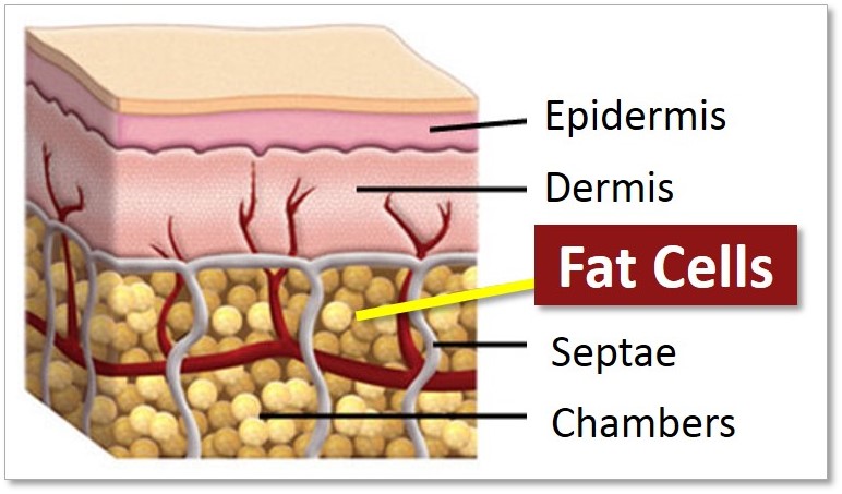
Adipose tissue, commonly referred to as fat tissue, is a highly vascularized, cellularly heterogeneous, and dynamic loose connective tissue. While characterized by its abundance of adipocytes—which specialize in storing lipid within a central lipid droplet—a substantial portion of its volume consists of the stromal vascular fraction (SVF). The SVF includes endothelial cells (lining the blood vessel networks within the tissue), immune cells, pericytes, and fibroblastic progenitor cells. The metabolic activity of these tissues hinges not only on the uptake and release of fatty acids but also on the secretion of endocrine factors, including adipokines (e.g., leptin, adiponectin) and cytokines (e.g., TNF-α, IL-6), which regulate systemic metabolism and inflammation.
Adipose distribution throughout the body is categorized into distinct depots, each with unique developmental origins and physiological impacts on body weight and metabolic health:
White Adipose Tissue (WAT): Composed primarily of white adipocytes (white fat), WAT arises from mesenchymal stem cells (MSCs) via PPARγ and C/EBPα pathways and serves as the body’s primary energy reservoir. Subcutaneous fat (SAT) and visceral adipose tissue (VAT) are the two main types. Visceral fat—specifically abdominal fat located around internal organs—is highly metabolically active; its expansion as excess adipose tissue is strongly linked to metabolic disease, diabetes, and heart disease.
Brown and Beige Adipose Tissue: Brown adipose tissue (brown fat) derives from a myogenic lineage shared with skeletal muscle, driven by PRDM16 and PGC-1α pathways. While present in humans, its activity declines with age and obesity. Beige adipocytes (beige fat) represent a recruitable population that can arise via de novo differentiation or the transdifferentiation of white adipocytes; their induction is a major focus of research for new obesity therapies.