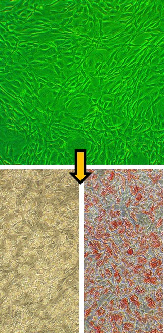
Human Preadipocytes: HPAd
Human Preadipocytes (HPAd) are mononucleated precursor cells isolated from the stromal vascular fraction of human adipose tissue.
Description
Human Preadipocytes (HPAd) — also referred to in the literature as adipocyte precursor cells — are mononucleated precursor cells isolated from the stromal vascular fraction of human adipose tissue. In their undifferentiated state, HPAd exhibit a fibroblast-like morphology and are heavily enriched or primed for adipogenic differentiation. Unlike completely multipotent human mesenchymal stem cells, these restricted precursor cells are already biased toward becoming mature, lipid-laden fat cells. To maintain this cell type in a proliferative, undifferentiated state in vitro, researchers utilize a specialized HPAd growth medium kit containing an optimized HPAd growth supplement. This culture setup allows the cells to expand for a limited number of passaged population doublings while preventing premature differentiation, enabling investigators to study preadipocyte proliferation dynamics prior to initiating the adipogenic cascade.
In the human body, these precursor cells reside within the connective tissue stroma of adipose tissue depots. They are commonly sourced from subcutaneous fat depots or specialized visceral depots, such as epicardial or omental fat. Within these tissue niches, HPAd are situated adjacent to mature adipose cells and capillaries, where they serve as a critical cellular reservoir for tissue expansion. While they share an initial spindle-shaped morphology with other stromal cell populations, their specific lineage commitment sets them apart. Upon receiving specific systemic or localized hormonal cues, these cells modify cell–matrix interactions to generate the functional lipid-storing units of the tissue.
The primary biological function of HPAd is to undergo adipogenesis to modulate fat mass expansion and maintain whole-body metabolic homeostasis. When exposed to an appropriate adipocyte differentiation medium in vitro, the cells undergo a dramatic morphological transition. They shift from a flat, fibroblastic shape into spherical, mature fat cells characterized by the accumulation of intracellular lipid droplets. This process is driven by complex adipogenesis signaling networks. Shifting biochemical inputs activate master transcriptional regulators, including peroxisome proliferator-activated receptor gamma (PPARγ) and CCAAT/enhancer-binding proteins (C/EBPs). This coordinated gene regulation program ultimately leads to the expression of the insulin receptor and other critical metabolic proteins, transforming the cell into an insulin-responsive, metabolically active endocrine unit capable of buffering circulating fatty acids.
In translational medicine and endocrinology, HPAd serve as a crucial in vitro model system for decoding the molecular biology of obesity, type 2 diabetes, and metabolic syndrome. Researchers leverage these cells to map adipogenesis signaling pathways and evaluate how novel therapeutic compounds affect lipid kinetics. For instance, investigators use HPAd to test how specific dietary bioactives or pharmacological candidates might intercept differentiation or limit the size of accumulating lipid droplets.
Furthermore, because dysfunctional fat accumulation drives progressive systemic disorders, HPAd models are widely deployed in co-culture configurations with immune cells to study paracrine tissue stress, macrophage recruitment, and the cellular blocks that stall insulin receptor signaling. By evaluating how these primary cultures respond compared to an established cell line, scientists can accurately isolate cell-type-specific mechanisms to advance adipocyte biology research and validate novel disease-modifying metabolic therapies.
- Interaction between infiltrating monocytes and adipocytes affects production of metalloproteinases and osteopontin, a proinflammatory cytokine, which ultimately leads to development of more adipose tissue and insulin resistance
- Adipocyte differentiation requires activation of Akt1 through the mTORC2-BSTA mechanism, leading to downstream suppression of FoxC2
- Instead of protecting from hyperglycemia-induced ER stress, like it does in other cells, in adipocytes a bioactive, endogenously produced compound taurine was shown to modulate the expression of adipokines under inflammatory conditions by inhibiting the STAT-3 signaling pathway (Kim, 2013a,c), and to inhibit differentiation of preadipocytes into adipocytes
- FGF21, which leads to reduction of body weight in animal models of obesity, was shown to act by modulating gene expression, phosphorylating Frs2a, Erk1/2, and Mypt1 and by increasing oxidative capacity of adipocytes via AMPK–SIRT1–PGC1a cascade
- Atrial natriuretic peptide (ANP) regulates lipid catabolism and reduce insulin resistance in HPAd by activating AMPK
- Adipocytokines are involved in normal pregnancy and pregnancy-induced hypertension
- Lotus seed extract exhibits anti-obesity and hypolipidaemic properties
- HPAd (along with our Human Dermal Fibroblasts) demonstrate the role of epigenetic modifications in increasing efficiency of iPS reprogramming
Details
| Tissue | Normal healthy human adipose tissue | |
|---|---|---|
| QC | No bacteria, yeast, fungi, mycoplasma, virus | |
| Character | Positive for lipid drops in cytoplasm & Oil Red O Staining, in Differentiation Med | |
| Bioassay | Attach, spread, proliferate in Growth Med | |
| Cryovial | 500,000 HPAd (2nd passage) frozen in Basal Medium w/ 20% FBS, 5% DMSO | |
| Kit | Cryovial frozen HPAd (802h/s-05a), Growth Medium (811-500), Subcultr Rgnt Kit (090K) | |
| Proliferating | Shipped in Gr Med, 3rd psg (flasks or plates) | |
| Doublings | Can be cultured 2 passages before differentiation into HAd | |
| Applications | Laboratory research use only (RUO). Not for human, clinical, diagnostic or veterinary use. |
Resources
FAQs
Need More Help?
Visit our comprehensive FAQ page for detailed answers to common questions.
Need More Help?
Visit our comprehensive FAQ page for detailed answers to common questions.
Primary Cell FAQs Powered by Bioz