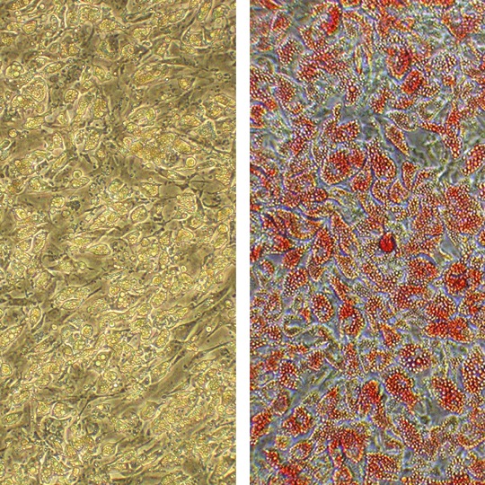
Human Adipocytes: HAd
Primary, mature, terminal-stage cells derived from the adipogenic differentiation of adipocyte precursor cells (human preadipocytes).
Description
Human Adipocytes (HAd) — alternatively referred to in scientific literature as mature fat cells or isolated adipocytes — are mature, terminal-stage cells derived from the adipogenic differentiation of adipocyte precursor cells (human preadipocytes). Unlike continuous cell lines that may lack human-specific metabolic networks, these cells capture native donor traits, rendering the mature human adipocyte a premier discovery platform for exploring adipocyte biology. Structurally, fully differentiated mature adipocytes shift away from a fibroblast-like precursor morphology to become spherical cells characterized by a large, unilocular central lipid droplet that displaces the cytoplasm and nucleus to the cell periphery. Functionally, each human adipocyte serves as the primary site for energy storage and functions as a critical endocrine organ that modulates whole-body metabolic homeostasis. To maintain their lipid-laden structure and support long-term cell survival in vitro, they are cultivated using specialized maintenance media.
In the human body, these cells are the primary structural components of adipose tissue, which is broadly categorized into subcutaneous fat (located directly beneath the skin) and visceral adipose tissue (enveloping the internal organs). The unique anatomical depot of origin, alongside donor variables such as age, sex, and body mass index (BMI), heavily dictates baseline fat cell size, gene regulation, and physiological responses. While the vast majority of human depots consist of white adipose tissue optimized for energy storage, specialized thermogenic depots also exist. In modern metabolic research, investigators frequently compare classic HAd profiles against specialized alternative lines, such as Human Brown Fat Preadipocytes (i-HBrPAd), to map the operational differences between energy-storing white fat and energy-expending brown or beige (brite) adipocytes.
The primary biological function of HAd is to govern systemic lipid metabolism by balancing lipogenesis (fat storage) and lipolysis (the enzymatic breakdown of triglycerides into free fatty acids and glycerol). This lipolytic cascade is precisely regulated by key sequential enzymes, primarily adipose triglyceride lipase (ATGL) and hormone-sensitive lipase (HSL), to control circulating fatty acid availability. Beyond serving as metabolic buffers, they secrete a broad range of signaling peptides, or adipokines (such as adiponectin and leptin), that coordinate peripheral insulin sensitivity and overall metabolic health.
However, chronic caloric excess leads to marked adipocyte hypertrophy, causing a significant expansion in individual adipocyte volume, overall fat cell size, and total fat mass. This pathological enlargement limits oxygen diffusion, triggering local hypoxia and endoplasmic reticulum (ER) stress. In these hypertrophic adipocytes, cellular stress can uncouple mitochondrial respiration and drive a pro-inflammatory secretory shift. While local hypoxia and altered mitochondrial respiration contribute substantially to inflammation, investigators recognize that other coordinated processes—such as extensive extracellular matrix remodeling and progressive pro-inflammatory macrophage recruitment—play major roles in driving overall adipose tissue dysfunction. Over time, these combined microenvironmental stressors compromise normal adipocyte function, contributing directly to chronic systemic inflammation, insulin resistance, and downstream weight gain.
In laboratory settings, HAd serve as a translationally rigorous human cell model for interrogating lipid kinetics, mapping endocrine signaling pathways, and validating therapeutics for metabolic disease and associated cardiovascular disease. Researchers utilize these cells to evaluate anti-obesity and hypolipidemic drug candidates, commonly measuring glycerol release from lipolysis alongside functional glucose uptake assays and glucose transporter 4 (GLUT4) membrane translocation monitoring. Additionally, because dysfunctional adipose tissue plays a prominent role in progressive metabolic disorders, type 2 diabetes, and metabolic syndrome, HAd are deployed in high-content screening pipelines to investigate how targeted weight loss interventions can reverse tissue stress and restore a favorable adipokine profile.
Furthermore, differentiated HAd are utilized in oncological research to examine adipocyte-tumor crosstalk. When provided as frozen cells in a cryopreserved vial, these primary cultures often reflect the baseline biological variation of their human origin. In postmenopausal breast cancer setups, investigators use these cells to study the localized upregulation of aromatase, helping map how local tissue-derived estrogen can drive estrogen receptor-positive tumor proliferation within the breast microenvironment.
Details
| Tissue | Normal healthy human adipose tissue | |
|---|---|---|
| QC | No bacteria, yeast, fungi, mycoplasma, virus | |
| Character | Positive for lipid drops in cytoplasm | |
| Cryovial | N/A | |
| Kit | N/A | |
| Cultured | Shipped at 1st passage in HAd Diff Med, after 5 days differentiation. Differentiated adipocytes do not divide. | |
| Doublings | N/A | |
| Applications | Laboratory research use only (RUO). Not for human, clinical, diagnostic or veterinary use. |
Resources
FAQs
Need More Help?
Visit our comprehensive FAQ page for detailed answers to common questions.
Need More Help?
Visit our comprehensive FAQ page for detailed answers to common questions.
Primary Cell FAQs Powered by Bioz