Human iPSC-Derived Hepatocytes: i-HH (Organoids)

Human iPSC-Derived Hepatocytes (i-HH) are specialized cells generated through the directed hepatic differentiation of an induced pluripotent stem cell (iPSC) line.

Quantity

Description

Human iPSC-Derived Hepatocytes (i-HH) — also classified as a specialized hepatocyte-like cell type or human induced pluripotent stem cells derived hepatic cells — are specialized cells generated through the directed hepatic differentiation of an induced pluripotent stem cell (iPSC) line. Traditionally, researchers have relied on human embryonic stem cells or primary human hepatocytes to study human liver tissue; however, primary liver cells tolerate cryopreservation poorly, display high donor-to-donor variability, and rapidly lose their phenotype in vitro. Conversely, immortalized cell lines lack normal xenobiotic metabolism. By taking advantage of a patient-specific pluripotent stem cell background, i-HH provide a scalable, non-malignant alternative. To transition these stem cells into functional liver cells, specialized cell differentiation protocols utilize sequential growth factor cocktails to drive definitive endoderm commitment, hepatic specification, and final maturation.

While these differentiated cells are generated in vitro, they are designed to recapitulate the cell biology and structural architecture of native hepatocytes within human liver tissue. In the human body, hepatocytes comprise the vast majority of the total liver mass, tightly organized into cords adjacent to vascular sinusoids lined with endothelial cells. To better mimic this intricate microenvironment, modern tissue engineering transitions flat monolayers into a three-dimensional (3D) liver organoid or a vascularized iPSC-derived liver organoids setup. During organoid formation within biomimetic matrices, i-HH can self-assemble alongside supporting non-parenchymal lineages — such as mesenchymal cells, endothelial cells, and hepatic stellate cells — to recreate the complex multicellular boundaries and structural polarization essential for long-term liver function.

The primary biological function of a mature hepatocyte is to maintain systemic metabolic homeostasis by executing critical biosyntheses and detoxification programs. Validated i-HH models are designed to capture this vital metabolic function, demonstrating the capability to synthesize and secrete plasma proteins like albumin, regulate glycogen storage, and clear toxic metabolic byproducts. A central metric of hepatic competence is their capacity for drug metabolism and xenobiotic metabolism, which relies on the coordinated gene expression and activity of phase I and phase II biotransformation networks.

Investigators should be cautious when claiming full equivalence to an adult mature hepatocyte. Many iPSC-derived hepatocytes often display a fetal-like phenotype, characterized by incomplete metabolic maturation and low baseline cytochrome P450 (CYP) expression compared with primary adult cells. Maturation can improve significantly with 3D culture and multi-lineage co-culture, and researchers routinely fix structures using 4% paraformaldehyde to execute multi-marker validation. This immunostaining for master transcription factors like HNF4A and surface receptors like ASGR1 is complemented by functional assays — such as active CYP assays, urea production, and bile acid transport — to accurately evaluate the functional status of the cells.

In preclinical drug discovery, toxicology, and regenerative medicine, i-HH setups serve as a translationally rigorous human-background platform. Safety pharmacologists deploy these cells in high-content screening pipelines to assess drug induced liver injury (DILI), evaluating how novel therapeutic candidates alter mitochondrial health, induce intracellular lipid accumulation, or trigger cholestasis. Because rodent animal models can fail to predict human-specific toxicity due to species differences in cytochrome P450 gene families, these human cells improve predictive validity.

Advanced multi-lineage liver organoid platforms are used to model progressive metabolic disorders like non-alcoholic fatty liver disease (NAFLD/MASH), map host-pathogen interactions for hepatitis viruses, and evaluate cell-encapsulation biomaterials designed to support long-term metabolic stability as bridging bio-artificial constructs for patients awaiting liver transplantation.

The following values represent typical ranges reported under specific assay conditions and vary based on assay format, sampling intervals, normalization methods, and vendor protocols:

Functional Biomarker Physiological Relevance Representative Primary Hepatocyte Range (Context-Dependent)
Albumin Secretion Major plasma protein essential for maintaining oncotic pressure and molecule transport. Normalized per volume over a standard timeline. 4 to 20 μg/mL per 24 h
Ammonia Detoxification The functional conversion of neurotoxic metabolic ammonia into water-soluble urea via the hepatic urea cycle. 48 to 255 nmol / 10^6 cells per 24 h
CYP3A4 Activity A principal cytochrome P450 of interest, responsible for metabolizing a large fraction of clinically prescribed drugs. 0.3 x 10^6 to 3.60 x 10^6 RFU (Relative Fluorescent Units; assay specific)
The extended viability of 3D i-HH organoids makes them an important tool across translational screening pipelines:

  • Drug-Induced Liver Injury (DILI) Screening: Unanticipated hepatotoxicity is a primary cause of late-stage pharmaceutical attrition. Because optimized i-HH organoids can maintain active Phase I (CYP450s) and Phase II conjugation enzymes over multi-week culture windows, safety toxicologists utilize them for chronic, repeated-dose toxicity testing to detect slow-onset or metabolite-mediated hepatotoxins.
  • Hepatic Metabolic and Infectious Disease Modeling: The patient-specific potential of HiPSCs allows for the generation of i-HH organoids carrying distinct genetic variants to model inherited metabolic disorders, non-alcoholic fatty liver disease (NAFLD/NASH), and glycogen storage pathologies. Additionally, they serve as a human platform for studying hepatitis virus replication kinetics. However, viral susceptibility depends heavily on the expression of specific entry receptors, co-factors, and native innate immune competence; not all i-HH organoids are fully permissive without further maturation or non-parenchymal co-culture.
Hepatocytes are essential for critical processes like metabolism, detoxification, protein synthesis, and innate immunity. A robust cellular model is required to study these hepatocyte processes, as well as liver development, toxicity, disease and more. However, current models have significant limitations.

Primary hepatocytes are inconvenient to isolate, do not tolerate cryopreservation, do not proliferate in culture, and senesce rapidly. Alternatively, cell lines like HepG2 can divide in vitro, but are cancer cells and do not display normal hepatocyte function. After about 1 week in standard cell culture format, cell lines like HepG2 becomes overgrown with necrotic colonies, and are no longer suitable for experiments. Furthermore, the majority of current hepatocyte models are 2-dimensional, a format that is unlikely to mimic the 3-dimensional niche that hepatocytes occupy in the body.

To address these liver research needs, Cell Applications now offers a breakthrough, first-of-its kind hepatocyte system. Using our DISCUS™ Technology to generate true three-dimensional tissue constructs, Cell Applications iPSC-derived Human Hepatocytes (i-HH) recapitulate hepatic development, generating 3D organoids with polarized structures capable of multiweek culture. The i-HH are well-characterized, and are demonstrated to express xenobiotic metabolizing enzymes, and demonstrate other key liver specific functions, including albumin secretion and ammonia detoxification.

i-HH demonstrates extended album secretion, urea production, and CYP3A4 activity, outperforming HepG2 in the DISCUS™ 3D system.


i-HH updated Albumin Urea CYP3A4 graphs 231205
i-HH updated Albumin Urea CYP3A4 graphs

Above graphs: Vital liver functions include production of albumin for molecule transport, urea from ammonia detoxification, and drug metabolizing enzymes like CYP3A4. As shown here, i-HH in the DISCUS™ 3D system possess all three activities. For reference, primary hepatocyte ranges are 4-20 ug/mL Albumin,2 48-255 nmol Urea,3 and 0.3-3.60E6 RFU CYP3A4 activity.4 Furthermore, i-HH outperforms HepG2 in both 2D monolayer (HepG2 -2D) and DISCUS™ 3D system (HepG2-3D) cultures. Adherent HepG2-2D cell cultures could only be cultured for one week before becoming overgrown.

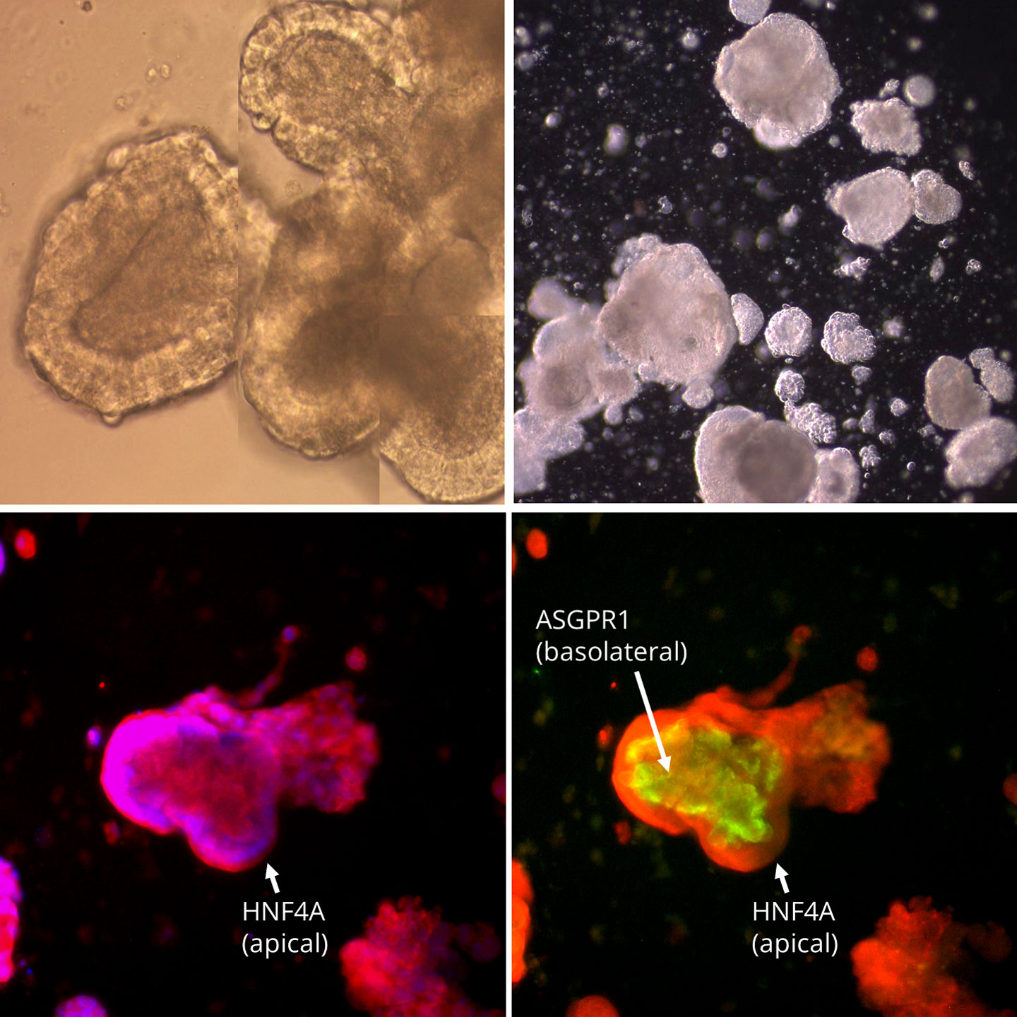
i-HH expresses liver markers HNF4A and ASPGR1, which are characteristic of functioning hepatocytes.1

Above Left: A 3D i-HH organoid stained with HNF4A (red) and ASPGR1 (green). The in-focus plane demonstrates a clear nuclear HNF4A signal on the apical exterior, and ASPGR1 signal on the basolateral interior. Above Right: HNF4A is a hepatocyte-specific transcription factor and ASPGR1 is a functional  hepatocyte marker. Cell Applications’ 3D i-HH, derived from two separate HiPSC lines (1709 and 1439), have 87% of the cells express double positive HNF4A/ASPGR1 markers for up to four weeks.

The i-HH Total Kit DISCUSTM system

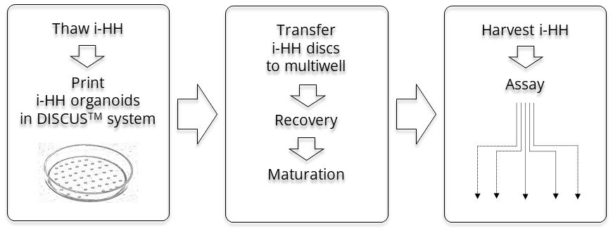
The HiPSC-derived Human Hepatocyte (i-HH) Total Kit from Cell Applications is a 3D cell culture model that utilizes the DISCUS™ gel system. The cell-specific DISCUS™ system of 3D discs address the challenges of anoxia and diffusion limits in 3D tissue engineering. Measuring 3mm across, and 600 microns deep at its thickest point, these versatile units need only common, inexpensive laboratory equipment to generate your 3D engineered tissue of choice. The i-HH Total Kit is an all-in-one package that includes cryopreserved i-HH, DISCUS™ gel kit, DISCUS™ dissociation solution, and optimized Recovery and Maturation Media that allow for the culture of the cells in a 3-D culture.

  1. Peters, et al. Development (2016) 1475-1481
  2. Lubberstedt, et al. J Pharm Tox Methods (2011) 59-63
  3. Donato, et al. Cell Transplantation (2008) 1211-1219
  4. Gramignoli, et al. Cell Tranplantation (2014) 1545-1556
Advanced Functional Characterization & Performance Data

To ensure maximum physiological relevance for drug screening, toxicity assays, and disease modeling, each batch of Human iPSC-Derived Hepatocytes (i-HH) undergoes rigorous functional and phenotypic validation in both 2D adherent and 3D organoid culture systems. Full experimental validation details can be reviewed in the technical resources below.

1. Phenotypic and Metabolic Validation
i-HH Biliary ducts and binucleate cells
Image (above): i-HH organoid explants on a coating of Laminin111/Collagen IV exhibit bile canuculi and binucleate cells typical of hepatocytes. Organoids grew in Recovery Media for four days before imaging.
Bile Duct Cholangiocyte System
Image (above): Overlay of flourescence with bright field
CFDA-2D: Biliary ducts are visible throughout a cluster of i-HH
Image (above): i-HH organoid explants on a coating of Laminin111/Collagen IV excrete fluorescent 5-CF which accumulates in bile canuculi. i-HH can form biliary ducts and has an active multidrug resistant-associated protein (MRP) transport system capable of excreting 5-carboxyfluorescein (5-CF). Organoids grew in Recovery Media for one week followed by Maturation Media for one week. Cells were then washed and incubated with 5 uM non-fluorescent 5-carboxyfluorescein diacetate (5-CFDA) for 1 hour at 37°C. Cells were washed three times with Maturation Media before imaging with a fluorescent microscope.
CFDA-3D: Biliary ducts visible throughout a 3D i-HH organoid
Images above and below (zoom): i-HH organoid embedded in 3D DISCUS system excretes fluorescent 5-carboxyfluorescein which accumulates in bile canuculi. i-HH can form biliary ducts and has an active multidrug resistant-associated protein (MRP) transport system capable of excreting 5-carboxyfluorescein (5-CF). Organoids incubated in Recovery Media for one week followed by Maturation Media for one week. Disks were then washed and incubated with 5 uM non-fluorescent 5-carboxyfluorescein diacetate (5-CFDA) for 1 hour at 37°C. Disks were washed three times 5 minutes each with Maturation Media, before imaging with a fluorescent microscope.
CFDA-3D zoom: Biliary ducts visible throughout a 3D i-HH organoid
      Image (left):
Glycogen Storage in i-HH is extremely heavy
Image (above): i-HH organoid explants on a coating of Laminin111/Collagen IV exhibit high levels of glycogen storage. Organoids grew in Recovery Media for one week followed by Maturation Media for one week. Cells were stained using Periodic-Acid Schiff for detection of glycogen according to manufacturer’s directions.
Phenylalanine Hydroxylase (PAH) and Transthyretin (TTR)
Image (above left): RT-PCR of adult liver (AD Liv) and i-HH demonstrates i-HH express PAH, the metabolic enzyme required to convert phenylalanine into tyrosine. Lack of this enzyme is the cause of phenylketonuria. Image (above right): RT-PCR of adult liver (AD Liv) and i-HH demonstrates i-HH express TTR (transthyretin), the transporter responsible for carrying thyroid hormones and retinol. i-HH organoids in the DISCUS system were incubated in Recovery Media for one week followed by Maturation Media for four weeks. Disks were dissolved directly in RLT buffer from Qiagen’s Rneasy kit and RNA isolated according to manufacturer’s directions. One microgram of RNA from each sample was then reverse transcribed and amplified.  

  • Hepatic Phenotype Validation: When cultured as organoid explants on a specialized Laminin-111/Collagen IV matrix, i-HH establish classic hepatic architecture, including the formation of visible bile canaliculi and a high frequency of binucleate cells typical of mature hepatocytes within 4 days of recovery.
  • Robust Glycogen Storage: Periodic Acid-Schiff (PAS) staining confirms exceptionally heavy intracellular glycogen accumulation within the organoid explants following a standard two-week maturation protocol.
  • Active Biliary Transport Systems: Functional Multidrug Resistance-Associated Protein (MRP/MRP2) transport systems are verified using a 5-CFDA efflux assay. The cells successfully internalize non-fluorescent 5-carboxyfluorescein diacetate (5-CFDA), cleave it into fluorescent 5-carboxyfluorescein (5-CF), and actively excrete it into interconnected canalicular networks across both 2D monolayer clusters and advanced 3D DISCUS-embedded organoids.
  • Key Mature Hepatic Gene Expression: Reverse Transcription Polymerase Chain Reaction (RT-PCR) of i-HH matured in the 3D DISCUS system demonstrates stable transcriptional expression profiles comparable to native adult liver tissue panels (AD Liv). This includes robust expression of Phenylalanine Hydroxylase (PAH, 205 bp), the metabolic enzyme required to convert phenylalanine into tyrosine, and Transthyretin (TTR, 100 bp), the plasma transport protein for thyroid hormones and retinol.

  2. Purity, Maturation Dynamics, & Infection Competency

iHH are HCV infection-competent: 94% are Occludin positive

  • HCV Infection Competence: Quantitative flow cytometry confirms that 94.3% of the i-HH population is positive for Occludin, a critical tight-junction protein and a primary co-receptor required for Hepatitis C Virus (HCV) entry and replication studies.


KRT7 versus Aquaporin – specificity for cholangiocytes
 

  • Regulated Cholangiocyte Differentiation Dynamics: Flow cytometry profiling of the biliary markers Cytokeratin 7 (KRT7) and Aquaporin-1 (AQP1) demonstrates a highly controlled maturation trajectory specific to cholangiocytes. Co-expression peaks significantly at Week 3 (86.5% double-positive) and naturally resolves by Week 4 (30.7%) as the population transitions into a stable, mature hepatic phenotype.

3. Structural Biliary Network Visualization

  • Co-Registration Imaging: High-resolution multi-modal visualization is achieved through the overlay of brightfield microscopy and fluorescence imaging, confirming the precise structural alignment and functional localization of active biliary excretion networks within the i-HH organoid structures.

Details

Source Derived from Human iPSC of a single donor
QC No bacteria, yeast, fungi, mycoplasma, virus
Bioassay Albumin secretion, Urea production, CY3A4 activity
Cryovial Approxiately 500,000 equivalents of i-HH (2nd passage) in freezing medium
Kit Cryovial frozen i-HH cells (i780-05), i-HH Recovery Medium Kit (i71103K-30), i-HH Maturation Medium Kit(i71105K-100), i-HH Metabolic Medium Kit (i71107K-30), DISCUS-111 i-HH Kit (DISC1053K), DISCUS Dissolution Reagent (DISC1050), Non-Stick Solution (1025-05)
Cultured Contact us for availability
Doublings Cells can be cultured for up to 3-4 weeks in DISCUSTM system. Cells do not divide, and cannot be passaged.
Applications Laboratory research use only (RUO). Not for human, clinical, diagnostic or veterinary use.
Instructions i-HH

Format: PDF

DOWNLOAD NOW
MSDS Cryopreserved Cells

Format: PDF

DOWNLOAD NOW

Resources

5 IMPORTANT CELL CULTURE RULES

Format: PDF

DOWNLOAD NOW

FAQs