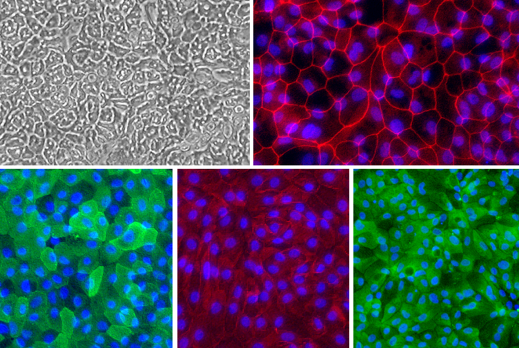
Human Colonic Epithelial Cell (HCnEpC) monolayer. Top left: Phase contrast. Top right (Red): ZO-1. Bottom left (Green): Villin. Bottom middle (Red): Na+/K+ ATPase. Bottom right (Green) pan-Cytokeratin. Blue: DAPI.
Human Colon Epithelial Cells: HCnEpC
Human Colon Epithelial Cells (HCnEpC) are the highly specialized epithelial cells that form the continuous mucosal barrier of the large intestine.
Description
Human Colon Epithelial Cells (HCnEpC) — extensively studied as human colonic epithelial cells or simply colonic epithelial cells — are the highly specialized epithelial cells that form the continuous mucosal barrier of the large intestine. Unlike continuous immortalized cell lines or cancer-derived models widely used in research (such as Caco-2, HT-29, or HCT116), true HCnEpC are human primary cells harvested from healthy or diseased human colonic tissue. Maintaining these primary cells requires specialized cell culture media, such as an optimized epithelial cell growth medium, to preserve their functional traits ex vivo. When cultivated under optimized conditions, these intestinal epithelial cells form a polarized monolayer characterized by functional tight junction networks. This structural baseline can be characterized by a marker panel including Villin (typically found on the apical membrane of enterocytic lineages), Na+/K+ ATPase and E-cadherin (basolateral markers), ZO-1 (tight junctions), and pan-Cytokeratin.
In the human body, these intestinal cells line the mucosal surface of the colon, organized into a series of deeply pocketed structural units called crypts. While the general architecture of the colonic mucosal lining shares structural similarities with the small intestine, the colonic intestinal epithelium is histologically unique because it features a flat surface topology entirely lacking villi. Within each crypt unit, a small, highly protected pool of intestinal stem cells resides at the crypt base. These multipotent stem cells continuously proliferate and differentiate as they migrate upward. The fully mature, differentiated epithelial cells eventually populate the flat luminal surface, where they directly interface with the complex gut microbiota and incoming dietary substrates.
The primary physiological function of the colonic epithelium is to act as a dynamic physical and immunological barrier that regulates nutrient and fluid absorption while preventing the translocation of harmful luminal contents. As a crucial front-line defense, these epithelial cells monitor the gut environment by recognizing bacterial antigens through surface receptors, initiating a highly calibrated immune response when pathogens are detected. Through the expression of dedicated response genes, they coordinate localized cytokine and chemokine networks to communicate with underlying immune cells. Furthermore, this dynamic barrier regulates homeostatic tissue regeneration, balancing rapid cell proliferation at the crypt base with programmed cell shedding at the luminal surface. To confirm the functional integrity of this barrier in vitro, researchers look for tight junction formation alongside functional assays such as transepithelial electrical resistance (TEER) or paracellular tracer flux.
In laboratory settings, HCnEpC serve as a vital primary cell translation tool in modern molecular biology and cell biologics. While researchers have historically relied on animal models utilizing mouse tissues, rat tissues, or tissues from rabbits and nonhuman primates, human-derived primary cells are essential to accurately recapitulate human-specific pathogenesis due to significant interspecies biological differences. Investigators utilize HCnEpC to study the cellular mechanics of widespread gastrointestinal disorders such as inflammatory bowel disease (IBD), Ulcerative Colitis, and colon cancer. Because primary colonic monolayers often have a limited lifespan in 2D culture (commonly about one week) without advanced support systems, researchers frequently adapt these cells into 3D biomimetic architectures. By manipulating specific cell culture media and environmental factors, adult stem cells can be grown as organoids and directed to become either stem cell enriched or enterocyte enriched, allowing for long-term evaluation of therapeutic mediators. These 3D systems permit researchers to analyze cellular cross-talk alongside co-cultured endothelial cells or mesenchymal stem cells to develop novel targeted treatments for chronic inflammatory and malignant colorectal diseases.
Details
| Tissue | Normal healthy human colon. | |
|---|---|---|
| QC | No bacteria, yeast, fungi, mycoplasma, virus. | |
| Bioassay | Attach, spread, in Culture Medium. | |
| Cryovial | 500,000 HCnEpC in Freezing Medium. | |
| Kit | Cryovial frozen HCnEpC (732Cn-05), Culture Med (716DC-50), Coating Solution (1024-05), Thawing Solution (716T-20). | |
| Cultured | Shipped in flasks or plates in medium. | |
| Doublings | Cells do not divide, and cannot be passaged. Cells can be cultured up to 7 days. | |
| Applications | Laboratory research use only (RUO). Not for human, clinical, diagnostic or veterinary use. |
Resources
FAQs
Need More Help?
Visit our comprehensive FAQ page for detailed answers to common questions.
Need More Help?
Visit our comprehensive FAQ page for detailed answers to common questions.
Primary Cell FAQs Powered by Bioz