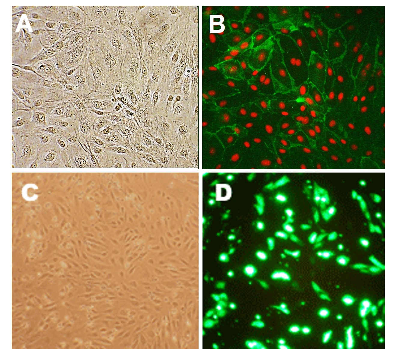
Human Lung Microvascular Endothelial Cells: HLMVEC
Human Lung Microvascular Endothelial Cells (HLMVEC) are highly specialized primary cells isolated from the dense alveolar microvasculature of the human lung.
Description
Human Lung Microvascular Endothelial Cells (HLMVEC) — alternatively designated as human primary pulmonary microvascular endothelial cells — are highly specialized primary cells isolated from the dense alveolar microvasculature of the human lung. Unlike immortalized cell lines, these primary cells have a limited proliferative capacity and will naturally undergo replicative senescence after a finite number of doublings; specialized cell culture media and an optimized endothelial cell growth media system can extend their lifespan. In vitro, HLMVECs form a characteristic cobblestone monolayer that can retain many native features under appropriate conditions, though investigators must monitor for transcriptional and phenotypic drift that can occur with extended passage and changing culture environments.
In the human body, these cells line the vast, intricate capillary networks within the peripheral parenchyma of the lung. This unique anatomical position places them at the critical blood-air interface, directly adjacent to alveolar epithelial cells to form a continuous, semi-permeable barrier optimized for efficient gas exchange. Within this native tissue architecture, HLMVECs constantly interact with passing blood cells and experience mechanical stretch from respiratory cycles. While standard academic and commercial cell lots focus on these human primary cells, comparative molecular biology workflows often isolate distinct resident cell type populations from the same tissue, such as pulmonary smooth muscle cell groups. Investigators may also utilize mouse or rat primary pulmonary endothelial cells (including C57BL6 mouse primary cells or rat primary cells), though distinct species differences in receptor expression and immune signaling profiles can limit direct translation to human disease states.
The primary biological function of HLMVECs is to maintain a tight, regulated endothelial permeability barrier while actively modulating systemic vascular tone, blood pressure, and local immune responses. These human microvascular endothelial cells highly express ACE and contribute substantially to angiotensin I → II conversion and bradykinin degradation. Additionally, in response to various inflammatory stimuli and some viruses—such as respiratory pathogens like influenza or SARS-CoV-2—these cells actively participate in host defense. They dynamically remodel their cell-to-cell junctional complexes (such as VE-cadherin and ZO-1) and upregulate surface adhesion molecules (like ICAM-1 and VCAM-1) to coordinate the tethering, rolling, and transendothelial migration of circulating leukocytes.
In advanced cell biologics and translational medicine, HLMVEC systems are widely leveraged as indispensable in vitro models to explore pulmonary injury cascades, model acute respiratory distress syndrome (ARDS), and screen therapeutic compounds. Investigators use these primary cell cultures to measure real-time changes in endothelial permeability during endotoxemic shock or to analyze cell-surface marker expression via flow cytometry. Because the extensive surface area and high blood flow of the lung capillary bed make it a frequent target for metastatic seeding, researchers utilize HLMVECs in tumor endothelial cell culture assays to study how circulating malignant cells attach to and extravasate through the lung endothelium.
Furthermore, these primary cell cultures serve as an essential biological baseline in regenerative medicine. They are frequently deployed alongside mesenchymal stem cells or pluripotent stem cells to investigate tissue vascularization, evaluate vector delivery using endothelial specific adenoviral overexpression tools, and test the safety profiles of novel cell biologics products before transitioning to complex in vivo validation models.
Details
| Tissue | Human lung | |
|---|---|---|
| QC | No bacteria, yeast, fungi, mycoplasma, virus | |
| Character | Factor VIII-related Ab, CD31 (PECAM-1), DiI-Ac-LDL uptake. S-HLMVEC are select HLMVEC lots that have been tested positive for VEGFR2 pathway activation following stimulation by VEGF. | |
| Bioassay | Attach, spread on AFS-coated surface, proliferate in Growth Med | |
| Cryovial | 500,000 HLMVEC (3rd passage) frozen in Basal Medium w/ 10% FBS, 10% DMSO | |
| Kit | Cryovial HLMVEC (540-05), Gr Med (111-500), Attchmnt Fctr Soln (123-100), Sbcltr Rgnt Kit (090K) | |
| Proliferating | Shipped in Tsfr Med, 4th psg (flasks or plates) | |
| Doublings | At least 15 | |
| Applications | Laboratory research use only (RUO). Not for human, clinical, diagnostic or veterinary use. |
Resources
FAQs
Need More Help?
Visit our comprehensive FAQ page for detailed answers to common questions.
Need More Help?
Visit our comprehensive FAQ page for detailed answers to common questions.
Primary Cell FAQs Powered by Bioz