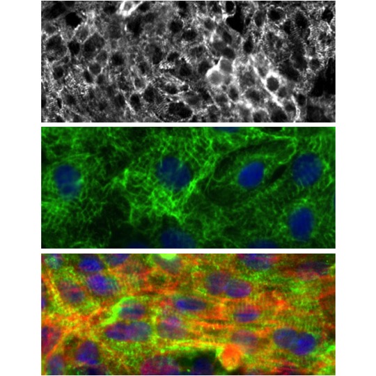
Rat Cardiomyocytes: RCm
Rat Cardiomyocytes (RCm) are specialized muscle cells isolated directly from normal rat heart tissue.
Description
Rat Cardiomyocytes (RCm) — frequently referred to as cardiac myocytes — are specialized muscle cells isolated directly from normal rat heart tissue. These cells serve as an invaluable in vitro model for investigating fundamental molecular biology pathways, the structural progression of heart failure, and the mechanisms of acute drug-induced cardiotoxicity. However, their physiological behavior and morphological stability in vitro are highly source-dependent.
Neonatal RCm typically exhibit robust, spontaneous, and synchronous rhythmic beating in culture, making them highly useful for baseline chronotropic assays. In contrast, isolated adult rat ventricular myocytes are generally quiescent unless electrically paced, and they present a greater technical challenge to maintain in long-term culture while preserving their classic rod-shaped morphology and delicate T-tubule networks.
When harvested via optimized cardiomyocyte isolation protocols, these isolated cells retain critical in vivo features such as mature sarcomeric alignment and functional excitation-contraction coupling. However, investigators must note that these complex properties are highly dependent on the isolation method and culture duration, as an adult cardiomyocyte will typically dedifferentiate over extended periods in vitro, leading to a loss of T-tubules and a decline in mature contractile features. Furthermore, because rodent electrophysiology and ion-channel expression differ fundamentally from human myocardium — such as shorter rodent action potential durations driven by distinct repolarizing currents—caution must be exercised when extrapolating translational readouts from an animal myocyte model to human clinical outcomes.
-
Mechanisms of Angiogenesis Inhibitor Cardiotoxicity: Primary cardiomyocyte platforms are widely used to identify secondary toxicities associated with modern oncology regimens. Researchers deploy RCm to examine the signaling cascades triggered by anti-cancer drugs that target vascular endothelial growth factor (VEGF) pathways. In these systems, drug exposure can induce mitochondrial dysfunction, altered energy production, and a loss of structural myofibrils, which helps model the mechanisms behind drug-induced clinical heart failure.
-
Investigating Drug-Induced Arrhythmias: Phenotypic screening in RCm helps identify compound-induced alterations in cardiac rhythmicity and electrical activity. For instance, while clinical cardiotoxicity and QT prolongation from antipsychotic drugs like thioridazine and mesoridazine are driven largely by human ether-à-go-go-related gene (hERG/IKr) potassium channel blockade in human patients, experimental studies in rodent single cell cultures indicate these compounds can also inhibit gap junction communication. These findings highlight that drug-induced electrical disturbances often involve multiple, interconnected cardiotoxic mechanisms.
-
Pathogen Toxins and Experimental Contractile Failure: Bacterial virulence factors are frequently evaluated in vitro to characterize toxin-induced myocardial injury. In specific primary experimental layouts, exposure to Clostridium difficile toxin B has been observed to rapidly disrupt the coordinated contractility and rhythmicity of neonatal RCm. Within this specific model system, the structural damage was shown to involve programmed cell death pathways, where the administration of a caspase-3 enzyme inhibitor helped preserve myocyte architecture and protect baseline contractile function.
-
GPCR Signaling and Ischemia-Reperfusion Models: Primary cultures are often deployed to study G-protein coupled receptor (GPCR) dynamics during simulated hypoxia and reoxygenation. In some experimental models, the Mas receptor has been shown to activate phospholipase C and drive inositol phosphate accumulation, contributing to simulated ischemia-reperfusion injury. While these downstream cascades can be highly context-dependent, this system has allowed researchers to screen for novel agonists and inverse agonists, with some studies demonstrating that inhibiting Mas function provides cardioprotective effects in isolated hearts and live mouse models.
-
Cardiac Regeneration and Advanced Diagnostic Imaging: Because the adult mammalian heart lacks a robust baseline capacity for cardiac regeneration, tracking therapeutic response and structural tissue remodeling is a major focus in heart disease research. RCm are utilized to evaluate how targeted growth factor regimens or transplanted stem cells influence local myocardial repair and cell viability. Concurrently, these cells serve as a standard validation matrix for testing novel diagnostic tools, such as positron emission tomography (PET) probes designed to improve high-resolution cardiac imaging.
Details
| Tissue | Normal healthy neonatal rat heart | |
|---|---|---|
| QC | No bacteria, yeast, fungi, mycoplasma | |
| Bioassay | Attaches on FBS-coated culture ware, Remains viable 2-4 days in Cltr Med | |
| Cryovial | N/A | |
| Kit | N/A | |
| Cultured | Shipped w/in 3 days after plating in Maint Med (flasks or plates) | |
| Doublings | N/A | |
| Applications | Laboratory research use only (RUO). Not for human, clinical, diagnostic or veterinary use. |
Resources
FAQs
Need More Help?
Visit our comprehensive FAQ page for detailed answers to common questions.
Need More Help?
Visit our comprehensive FAQ page for detailed answers to common questions.
Primary Cell FAQs Powered by Bioz