Human Aortic Smooth Muscle Cells: HAOSMC
Human Aortic Smooth Muscle Cells (HAOSMC) are the primary cellular component of the medial layer of the human aorta.
Description
Human Aortic Smooth Muscle Cells (HAOSMC) are the primary cellular component of the medial layer of the human aorta, playing a central role in maintaining structural integrity, vascular tone, and homeostatic blood pressure within the macrovasculature. As a highly specialized cell type, these vascular smooth muscle cells (VSMC) are not terminally differentiated; instead, they retain a remarkable capacity for dynamic phenotypic switching.
In a healthy aortic wall, HAOSMC maintain a quiescent, contractile phenotype characterized by the high expression of contractile apparatus proteins. However, in response to vascular injury, mechanical stress, or inflammatory stimuli, they can transition into an active, proliferative phenotype (also referred to as a synthetic or secretory state). This phenotypic plasticity is a foundational driver of vascular disease, making HAOSMC an essential framework for studying the cellular mechanisms of intimal hyperplasia, cellular hypertrophy, medial degeneration, and vascular calcification.
In the laboratory, establishing a cell culture using authentic primary cells isolated directly from normal human aorta tissue provides a superior alternative to continuous cell line options, which frequently display altered signaling kinetics and modified phenotypic stability. When maintained in a specialized SMC medium, primary HAOSMC retain their native responsiveness to mechanical stretch, extracellular matrix (ECM) composition, and local cytokine signaling. This makes them highly valued reagents for modern vascular disease research, enabling investigators to map the molecular mechanics behind occlusive vasculopathies, evaluate advanced endovascular stent coatings, and optimize targeted therapeutic delivery systems.
-
Phenotypic Switching and Atherosclerosis: A major focus of vascular smc research is dissecting the molecular triggers that govern the transition from a contractile to a proliferative phenotype. During the development of atherosclerosis and post-angioplasty stent restenosis, medial HAOSMC downregulate contractile markers and migrate into the intima. Here, they proliferate rapidly, secrete large volumes of extracellular matrix, and interact closely with infiltrating macrophages and overlying endothelial cells to form the fibrous cap of atherosclerotic plaques, driving the structural progression of arterial narrowing.
-
Aortic Dissection and Severe Arterial Disease: Degeneration of the vascular media is a foundational feature of life-threatening macrovascular events, such as thoracic aortic aneurysms and aortic dissection. The mechanisms driving these conditions are multifactorial, involving profound cell loss, extensive ECM proteolysis, and chronic hemodynamic strain. Furthermore, arterial disease pathways are heavily influenced by genetic mutations — such as those altering fibrillin-1 or elements of the TGF-β signaling pathway — which disrupt the structural integrity of the aortic wall. Primary HAOSMC models allow researchers to investigate how these genetic and mechanical defects compromise wall stability, providing a translational model to evaluate novel pharmacological interventions.
-
Vascular Calcification and Complex Transdifferentiation: In patients suffering from diabetes or chronic kidney disease, HAOSMC are exposed to persistent metabolic stress that can stimulate an osteogenic transdifferentiation program. Investigators utilize primary cultures to study vascular calcification, tracing how mineral deposition is regulated by a complex network of signaling pathways (including BMPs, Runx2, and Wnt signaling). Within this multifaceted pathological cascade, altered regulation of intracellular calcium channels represents one critical factor among many that disrupt mineral homeostasis and increase arterial stiffness.
-
Advanced Stent Technology and Microfluidic Arrays: Evaluating the compatibility and therapeutic efficacy of endovascular devices requires high-fidelity physiological testing. By seeding primary HAOSMC onto specialized surfaces or inside advanced microfluidic layouts, researchers can analyze cell attachment and plating efficiency on novel biomaterials. These microphysiological setups routinely apply fluid shear stress or cyclic stretch to study how mechanical forces modulate cell proliferation, migration, and drug-elution kinetics from advanced stent alloys.
-
Regenerative Medicine and Stem Cell Lineage Comparison: Understanding the differentiation limits of vascular wall components is critical for tissue engineering and vessel replacement therapies. Researchers frequently contrast the functional kinetics and baseline secretome of primary HAOSMC against smooth muscle cells derived from multi-lineage stem cells (such as induced pluripotent stem cells or bone marrow-derived mesenchymal populations), often utilizing specialized stem cell differentiation kits. These comparison models help benchmark the maturity, contractile function, and safety of cell sources intended for bioengineered vascular grafts.
- Cytokines and growth factor signaling pathways implicated in the molecular regulation of smooth muscle cell proliferation, migration, and overall vascular function
- Hyperglycemia-related risk factors for atherosclerosis in diabetes patients, as well as effects of ethanol on vascular calcification
- ECM deposition and its role in cardiovascular health, repair of damaged vasculature and successful tissue engineering
- Mechanisms and effects of mechanoregulation on proliferation and function of smooth muscle cells
- Advanced stent technology, including novel surface materials
- Drug and gene delivery systems
- Restenosis and other occlusive vasculopathies
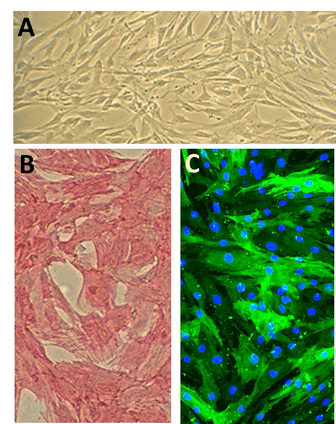
Characterization: Positive for smooth muscle cell specific alpha-actin expression.
Details
| Tissue | Normal healthy human aorta | |
|---|---|---|
| QC | No bacteria, yeast, fungi, mycoplasma, virus | |
| Character | Smooth muscle specific α-actin positive | |
| Bioassay | Attach, spread, proliferate in Growth Med | |
| Cryovial | 500,000 HAOSMC (2nd passage) frozen in Basal Medium w/ 10% FBS, 10% DMSO | |
| Kit | Croyvial frozen HAOSMC (354-05a), Growth Medium (311-500), Subcltr Rgnt Kit (090K) | |
| Proliferating | Shipped in Gr Med, 3rd psg (flasks or plates) | |
| Doublings | At least 16 | |
| Applications | Laboratory research use only (RUO). Not for human, clinical, diagnostic or veterinary use. |
Resources
FAQs
Need More Help?
Visit our comprehensive FAQ page for detailed answers to common questions.
Need More Help?
Visit our comprehensive FAQ page for detailed answers to common questions.
Primary Cell FAQs Powered by Bioz