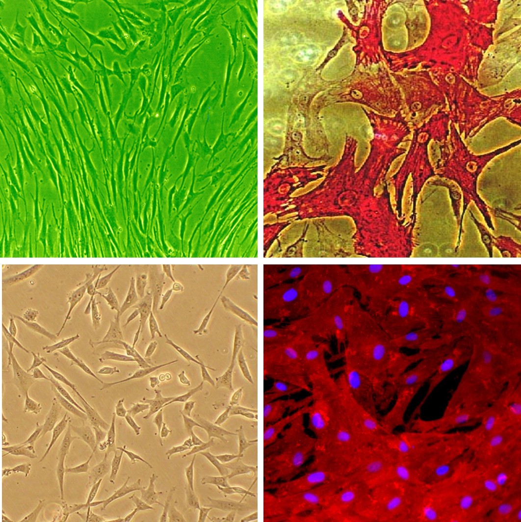
Human Coronary Artery Smooth Muscle Cells: HCASMC are isolated from two major vessels that supply the heart with blood. TOP: Phase-contrast microscopic image under green light (L), Differentiated HCASMC stain positive for smooth muscle cell-specific alpha-actin expression (R). BOTTOM: Phase contrast (L) and
...Human Coronary Artery Smooth Muscle Cells: Plaque: HCASMC-q
Human Coronary Artery Smooth Muscle Cells from Atherosclerotic Plaque (HCASMC-q) are primary cells derived from plaque tissue within human coronary arteries.
Description
Human Coronary Artery Smooth Muscle Cells from Atherosclerotic Plaque (HCASMC-q) are primary cells derived from plaque tissue within human coronary arteries. While standard primary smooth muscle cells from healthy tissue serve as a baseline control, HCASMC-q provide a disease-altered in vitro model.
In a healthy blood vessel, contractile vascular smooth muscle cells maintain structural integrity and vascular tone. However, within an active lesion, these cells undergo a dramatic shift, downregulating hallmark contractile markers like alpha smooth muscle actin (α-SMA) to take on highly plastic, non-contractile phenotypes.
Rather than reverting to a single alternative cell type, fate-mapping and single-cell sequencing show that these vascular smc populations exhibit profound heterogeneity. Depending on the local microenvironment, subsets of these cells have been shown to transition toward macrophage-like, fibroblast-like (or myofibroblast-like), or osteogenic phenotypes.
These cells are plaque-derived, originating within the epicardial coronary arteries—the main vessels supplying oxygenated blood to the myocardium. This diseased microenvironment is distinct from healthy tissue types. It involves a complex interplay between the tunica media and the subendothelial space, where damaged endothelial cells allow lipids to infiltrate the vessel wall.This specific vascular pathology is unique compared to non-vascular tissues, such as skeletal muscle, or other distinct stem cell niches like the bone marrow, the umbilical cord, or the central nervous system containing neural stem cells.
HCASMC-q function as a specialized model to study the chronic, multi-lineage cellular transformations that drive advanced cardiovascular disease and precipitate clinical events like an acute myocardial infarction (heart attack).
1. Neointimal Expansion and Plaque Remodeling
During atherosclerotic plaque formation, modified smooth muscle cells migrate from the media into the intima. This migration, combined with localized cell proliferation, drives the cellularity of the expanding lesion, gradually narrowing the arterial lumen.
2. Lipid Handling and Defective Clearance
Within atherosclerotic lesions, shifted smooth muscle cells take up excess lipid molecules, developing a foam-cell phenotype that resembles macrophages but lacks effective clearance capabilities. Over time, persistent lipid accumulation and localized stress trigger cell apoptosis (programmed cell death). When these cells die, they release cell debris, lipids, and necrotic material into the core, expanding the necrotic zone and destabilizing the plaque.
3. Fibrous Cap Dynamics vs. Vascular Calcification
The balance between matrix synthesis and degradation determines plaque stability. Transformed smooth muscle cells can take on a fibroblast-like role, producing a collagen-rich fibrous cap that insulates the thrombogenic core from blood flow.
Conversely, chronic inflammatory signaling can induce an osteogenic shift. In this state, the cells express bone-related protein networks, releasing matrix vesicles that nucleate calcium deposits. This active mineralization alters the mechanical compliance of the arterial wall, directly influencing whether a plaque remains stable or becomes vulnerable to rupture.
Because public health heavily prioritizes therapeutic interventions for advanced coronary artery disease, HCASMC-q serve as a highly translational, human-derived platform for preclinical testing.
Disease Modeling & Phenotypic Assays
Researchers isolate these donor specific cells to evaluate how chronic, in vivo disease programming persists during in vitro propagation. In the laboratory, scientists process these cultures into a concentrated cell pellet to perform Western blotting, qPCR, and mass spectrometry, tracking how specific gene programs deviate from healthy controls.
Culture Optimization & In Vitro Workflows
To preserve their delicate, disease-specific profiles without introducing confounding variables, investigators often utilize advanced xeno-free cell culture workflows. By testing these primary cells with specialized attachment factors and defined medium formulations, researchers can map out the precise molecular signals that drive smooth muscle cells toward a protective, fibrous phenotype rather than a destructive, calcifying state. Understanding how these pathways differ from true stem cells or validated fibroblasts helps scientists identify unique therapeutic targets to stabilize vulnerable plaques in patients.
Human Coronary Artery Smooth Muscle Cells (HCASMC) provide an excellent model system to study all aspects of cardiovascular function and disease, especially those related to mechanisms of hyperplasia and hypertrophy of intimal smooth muscle cells leading to vascular occlusion in atherosclerosis and stent restenosis.
HCASMC from Cell Applications, Inc. have been utilized in a number of research studies, for example, to:
- Study signaling pathways regulating smooth muscle differentiation and chronic inflammation of arterial wall that leads to artherosclerosis
- Demonstrate that STAT-1 and STAT-3 regulate VEGF production in smooth muscle cells by having opposing effects on HIF-1α expression
- Examine the mechanisms of hypoxia and reoxigenation injuries in by demonstrating increased production of ROS and inflammatory cytokines, and further showing that DHA is not beneficial in this type of injuries
- Investigate (by also using Human Internal Thoracic Artery Smooth Muscle Cells obtained from Cell Applications, Inc.), the gene expression differences between smooth muscle cells from different arteries, underlying their differential response to injuries and proliferation stimuli
- Suggest the hypermethylation of SOCS3 gene as the connection between TNF-α and IGF-1 released in response to mechanical injury during coronary intervention, and the induction of cytokines leading to intimal hyperplasia and restenosis
- Develop a novel VEGFR/MET-targeted inhibitor with improved antitumor efficacy and decreased toxicity
- Investigate novel therapies and drug combinations to achieve optimal target selectivity
- Generate elastic scaffolds for tissue engineering and novel treatment strategies to prevent stent restenosis by designing new materials, or drug therapies to preferentially inhibit smooth muscle cell growth
Characterization: positive for smooth muscle cell specific alpha-actin expression
Details
| Tissue | Human coronary artery from donor with plaque | |
|---|---|---|
| QC | No bacteria, yeast, fungi, mycoplasma, virus | |
| Character | Smooth muscle specific α-actin positive | |
| Bioassay | Attach, spread, proliferate in Growth Med | |
| Cryovial | 500,000 HCASMC-q (2nd passage) frozen in Basal Medium w/ 10% FBS, 10% DMSO | |
| Kit | Cryovial frozen HCASMC-q (350q-05a), Growth Medium (311-500), Subcltr Rgnt Kit (090K) | |
| Proliferating | Shipped in Gr Med, 3rd psg (flasks or plates) | |
| Doublings | At least 16 | |
| Applications | Laboratory research use only (RUO). Not for human, clinical, diagnostic or veterinary use. |
Resources
FAQs
Need More Help?
Visit our comprehensive FAQ page for detailed answers to common questions.
Need More Help?
Visit our comprehensive FAQ page for detailed answers to common questions.
Primary Cell FAQs Powered by Bioz