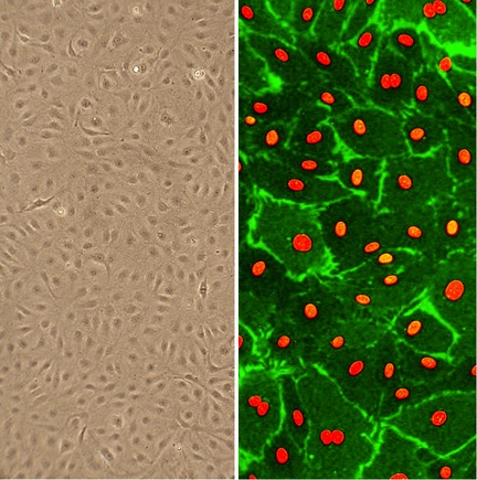
(Cllick to Enlarge) Human Dermal Lymphatic Microvascular Cells (HDLMVEC) in culture (L). HDLMVEC immunolabeled for CD31/PECAM (green); nuclei are visualized with PI (red) (R).
Human Dermal Lymphatic Microvascular Endothelial Cells: HDLMVEC
Human Dermal Lymphatic Microvascular Endothelial Cells (HDLMVEC) are primary cells isolated from the specialized network of lymphatic capillaries within normal human dermis.
Description
Human Dermal Lymphatic Microvascular Endothelial Cells (HDLMVEC) are primary cells isolated from the specialized network of lymphatic capillaries within normal human dermis. Unlike immortalized cell line alternatives, which often undergo phenotypic drift and lose complex in vivo behaviors, these human lymphatic endothelial cells retain their distinct physiological properties and morphology in vitro. Depending on the specific isolation protocol and application, these cells are typically derived from normal human skin samples provided by selected donors.
Because primary lymphatic endothelial cells require specific growth conditions to prevent dedifferentiation, they are cultured using a specialized endothelial cell medium or an optimized Microvascular Endothelial Cell Growth Medium, depending on the downstream assay requirements. Rather than establishing quality on continuous cell proliferation alone, standard endothelial quality control includes verification against validated tested markers. Characterization includes confirming the expression of the vascular endothelial growth factor receptor 3 (VEGFR3), along with endothelial markers such as Factor VIII-related antigen and CD31 (PECAM-1), alongside functional verification via fluorescently labeled acetylated low-density lipoprotein (DiI-Ac-LDL) uptake assays.
In vivo, these cells form the innermost lining of the initial lymphatic capillaries located within the dermal layer of human skin. The dermal microenvironment represents a complex matrix where these capillaries are situated alongside blood vessels, working in parallel to maintain peripheral fluid balance. Within this tissue architecture, lymphatic endothelial cells interact closely with surrounding dermal fibroblasts and migrating immune cells.
While they share features with other microvascular endothelial cell systems, lymphatic endothelial cells possess structural adaptations unique to their drainage functions. Unlike the tightly apposed cells lining blood capillaries, the HDLMVEC in initial lymphatic vessels are connected by discontinuous, button-like junctions and lack a continuous basement membrane. This overlapping layout forms a series of specialized micro-valves that make the initial lymphatic vessel highly permeable to fluid, macromolecules, and leukocytes.
The primary function of HDLMVEC is to maintain tissue homeostasis by regulating the drainage and transport of interstitial fluid, proteins, and lipids from the extracellular matrix back to the blood circulation. By dynamically modulating their surface adhesion molecules and junctional permeability, these cells actively participate in regional cell signaling and immune cell trafficking.
During tissue injury, localized signaling pathways stimulate these primary cells to initiate lymphangiogenesis — the growth of new lymphatic vessels — which is an essential step in physiological skin wound healing. However, this migratory and proliferative machinery can be hijacked under pathological conditions. In oncology, tumor lymphangiogenesis provides an important route for tumor metastasis, enabling tumor cells to enter the lymphatic drainage system and migrate to regional lymph nodes and distant organs.
In biomedical research, HDLMVEC serve as a useful model system to investigate the cellular mechanisms underlying vascular development, inflammatory trafficking, and oncology. They are widely utilized in cell proliferation, migration, and capillary tube-formation assays to map the specific signaling pathways that control lymphatic growth and barrier function. Scientific references frequently document the co-culture of HDLMVEC alongside complementary cell types, such as Human Umbilical Vein Endothelial Cells (HUVEC) or dermal fibroblasts, to study the roles of specific transcription factors (like Prox1 and FOXC2) during tumor lymphangiogenesis associated with squamous cell carcinoma progression.
Beyond cancer biology, these primary cells are widely used as advanced cell culture models to evaluate the safety and efficacy of topical therapeutics, study the pathobiology of lymphedema, and analyze the regulatory cross-talk between stem cells and the microvascular niche during tissue regeneration. Their ability to accurately mimic the specialized behavior of human dermal lymphatics makes them vital to modern microvascular and dermatological research.
Human Dermal Lymphatic Microvascular Endothelial Cells (HDLMVEC) are isolated from lymphatic capillaries of normal human dermis. The lymphatic system interacts closely with the blood vascular system in maintaining tissue homeostasis. It plays a vital role in the body by regulating the immune system, transporting interstitial fluid, proteins and fat to the blood circulatory system. Lymphangiogenesis is also implicated in the metastatic process. Although lymphatic microvascular endothelial cells have many properties in common with blood microvascular endothelial cells, they also have unique functions due to their structural features. Lymphatic capillaries have thin and discontinuous base membranes, and lymphatic endothelial cells, unlike their blood vessel counterparts, are not as tightly connected with each other, rendering the capillaries highly permeable.
HDLMVEC, together with Human Umbilical Vein Endothelial Cells, both from Cell Applications, Inc., have recently been used in a study demonstrating the key role of Prox1 and FOX C2 proteins in angiogenesis and lymphangiogenesis associated with oral squamous cell carcinoma progression.
Characterization: They express VEGFR3, and respond to activation by VEGF-C (see Figure on left).
Details
| Tissue | Normal healthy human lymphatic capillaries from dermis | |
|---|---|---|
| QC | No bacteria, yeast, fungi, mycoplasma, virus | |
| Character | Factor VIII-related Ag, CD31 (PECAM-1), DiI-Ac-LDL uptake | |
| Bioassay | Attach, spread, proliferate in Growth Med (AFS-coated surface) | |
| Cryovial | 500,000 HDLMVEC (3rd psg) frozen in Basal Medium w/ 10% FB, 10% DMSO | |
| Kit | Cryovial of HDLMVEC (100L-05a), Gr Med (111-500), Attchmnt Fctr Soln (123-100), Subcltr Rgnt Kit (090K) | |
| Proliferating | Shipped in Tsfr Med, 4th psg (flasks or plates) | |
| Doublings | At least 12 | |
| Applications | Laboratory research use only (RUO). Not for human, clinical, diagnostic or veterinary use. |
Resources
FAQs
Need More Help?
Visit our comprehensive FAQ page for detailed answers to common questions.
Need More Help?
Visit our comprehensive FAQ page for detailed answers to common questions.
Primary Cell FAQs Powered by Bioz