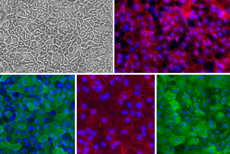
Human Intestinal Epithelial Cell (HInEpC) monolayer. Top left: Phase contrast. Top right (Red): ZO-1. Bottom left (Green): Villin. Bottom middle (Red): Na+/K+ ATPase. Bottom right (Green) pan-Cytokeratin. Blue: DAPI.
Human Intestinal Epithelial Cells: HInEpC
Human intestinal epithelial cells (HInEpC) are human intestinal epithelial cells organized in vivo into a single layer of cells forming crypts and villi.
Description
Human intestinal epithelial cells (HInEpC) — frequently referred to in research as human intestinal epithelial cells or simply HInEpC — are organized in vivo into a single layer of cells forming crypts and villi. Recognized as one of the most rapidly self-renewing tissues in adult mammals, this intestinal cell monolayer performs the primary functions of digestion, water and nutrient absorption, and mucosal defense. Within the crypts, intestinal stem cells continuously divide to generate transit-amplifying cells, which typically undergo a series of divisions—often generalized as 4–5 times, though this varies by species and specific intestinal region — before terminally differentiating into specialized epithelial cells.
These mature cell types include nutrient-absorbing enterocytes, goblet cells that secrete a protective mucus blanket, enteroendocrine cells that regulate metabolic homeostasis, and paneth cells that reside in the crypt bases to secrete antimicrobial peptides. Following terminal differentiation, cells migrate along the crypt-villus axis toward the tip of the villus, where they undergo spontaneous apoptosis and are shed into the gut lumen; this transit process takes an approximate or typical lifespan of a few days, depending significantly on the species and gut segment.
In the laboratory, establishing a cell culture using human primary cells — such as primary cells from the small intestine or human colonic epithelial cells — provides outstanding resources for investigating native gastrointestinal physiology. While an immortalized or transformed cell line like caco 2 cells is a transformed colorectal adenocarcinoma line used for specific functional assays like in vitro permeability testing, it carries known limitations. These include significant heterogeneity between strains and laboratories, altered enzyme or transporter expression profiles, and a lack of the complete cellular diversity found in vivo.
Consequently, primary cell platforms, human intestinal epithelial cell HInEpC monolayers, and advanced organoid cultures are frequently utilized as higher-fidelity models for specialized endpoints. Though the absolute physiological accuracy of these primary models still depends heavily on culture conditions — such as the presence of supportive stromal cell types, immune cells, or microfluidic flow — when paired with a specialized epithelial cell growth medium, these human intestinal cells form polarized monolayers that serve as invaluable assets for modern drug development and predictive toxicology.
-
Gastrointestinal Infection and Pathogen Barriers:
Researchers deploy normal colonic epithelial cells and small intestinal primary cultures to study host-pathogen interactions in real time. These models allow investigators to map how enteric bacteria, viruses, or fungi interact with the apical surface of the enterocyte. Primary systems are critical for observing how goblet cells alter mucus production and how tight junctions dynamically restrict pathogen translocation across the epithelial barrier.
-
Inflammatory Bowel Disease (IBD) and Cytokine Signaling:
In chronic inflammatory disorders such as Crohn’s disease and ulcerative colitis, the epithelial barrier is compromised by a cascade of inflammatory mediators. Primary human intestinal cells allow researchers to study epithelial breakdown, localized oxidative stress, and the failure of crypt-regulated regeneration. Investigators utilize these platforms to evaluate how therapeutic compounds can mitigate barrier porosity and accelerate mucosal healing in the colon.
-
Colorectal Oncogenesis and Tumor Initiation:
Primary epithelial models provide a crucial baseline for studying genetic instability and the initiation of colorectal malignancies. By comparing normal colonic epithelial cells against mutated lineages, investigators can analyze how oncogenic transformations disrupt the homeostatic balance between crypt-based stem cells and differentiated cells. This provides a clear window into how early-stage lesions bypass normal cell-cycle checkpoints.
-
Regenerative Medicine and Stem Cell Synergy:
Understanding the native niche of intestinal stem cells is central to developing cell-based therapies for short bowel syndrome and severe mucosal damage. Researchers routinely utilize organoid cultures to study the paracrine signaling networks between the epithelium and underlying stromal support. This includes analyzing how mesenchymal stem cells or localized stem cell populations secrete essential signaling factors to drive epithelial repair, proliferation, and architectural homeostatic balance.
When maintained in an optimized epithelial cell growth medium, primary HInEpC populations transition into a highly organized, functional monolayer. While structural polarization is broadly confirmed via general epithelial markers, researchers routinely employ a combined panel of general and lineage-specific markers for granular validation of individual cell subtypes:
-
Structural Domains and Polarity Markers:
-
Villin: An actin-modifying protein localized to the microvillar brush border to verify the apical domain.
-
Na+/K+ ATPase: Utilized to confirm the targeted localization of basolateral transport machinery.
-
Zonula Occludens-1 (ZO-1): Establishes the continuous, peripheral tight junction network sealing adjacent cells.
-
Pan-Cytokeratin: Used as a broad baseline stain to confirm the preservation of intermediate filament frameworks across all cells of epithelial origin.
-
-
Granular Lineage-Specific Subtype Markers:
-
Lgr5: Deployed to identify and track active intestinal stem cells within the crypt base.
-
MUC2 (Mucin-2): Utilized for the explicit identification of mucus-secreting goblet cells.
-
Chromogranin A: Expressed specifically to distinguish hormone-secreting enteroendocrine cells.
-
Lysozyme: Targeted to identify specialized, alpha-defensin-producing paneth cells.
-
Details
| Tissue | Normal healthy human intestine. | |
|---|---|---|
| QC | No bacteria, yeast, fungi, mycoplasma, virus. | |
| Bioassay | Attach, spread, in Culture Medium. | |
| Cryovial | 500,000 HInEpC in Freezing Medium. | |
| Kit | Cryovial frozen HInEpC, Culture Med (716DC-50), Coating Solution (1024-05), Thawing Solution (716T-20). | |
| Cultured | Shipped in flasks or plates in medium. | |
| Doublings | Cells do not divide and cannot be passaged. | |
| Applications | Laboratory research use only (RUO). Not for human, clinical, diagnostic or veterinary use. |
Resources
FAQs
Need More Help?
Visit our comprehensive FAQ page for detailed answers to common questions.
Need More Help?
Visit our comprehensive FAQ page for detailed answers to common questions.
Primary Cell FAQs