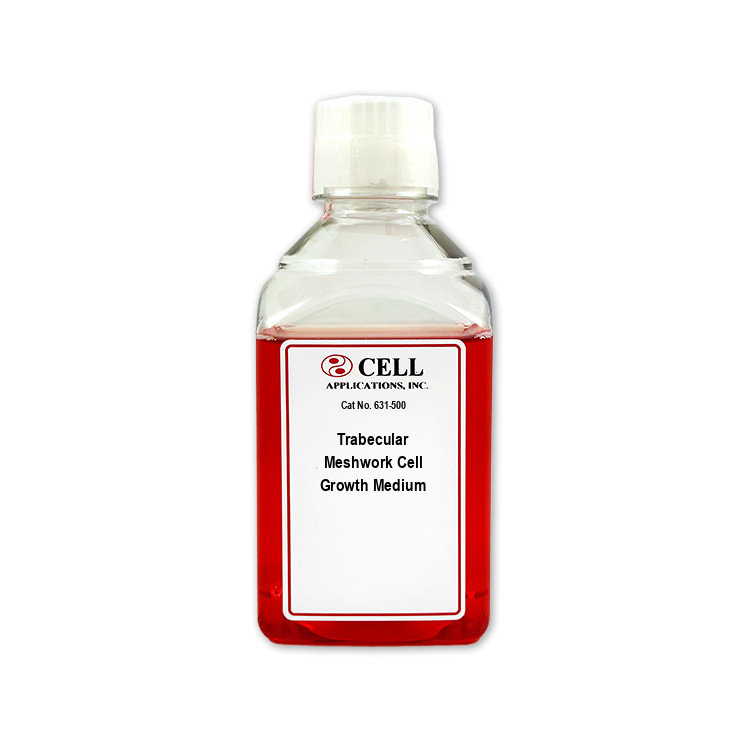
Human Trabecular Meshwork Cell Media, optimized for sterile, in vitro culture of HTMC
Human Trabecular Meshwork Cell Media
HTMC Growth Medium: All-in-one ready-to-use
Type: Growth Medium
Size: 500 ml
Price: $143.00
Description

Human Trabecular Meshwork Cell Media is an optimized growth formulation developed to support the attachment, viability, proliferation, and culture performance of primary human trabecular meshwork cells (HTMC) in vitro. The medium provides nutritional and biochemical components intended to support cell health, morphology, and reproducible culture performance during experimental manipulation. Because primary HTMC phenotype and function can vary with donor, passage number, substrate, and culture conditions, researchers should independently evaluate relevant phenotypic and functional markers for their specific application.
The trabecular meshwork is located within the iridocorneal angle of the anterior segment of the eye and is conventionally divided into three structural regions:
- Uveal Meshwork: The innermost region, adjacent to the anterior chamber, containing relatively widely spaced trabecular beams lined by trabecular meshwork cells.
- Corneoscleral Meshwork: The intermediate region, composed of multiple layers of connective-tissue beams containing collagenous and elastic elements and lined by trabecular meshwork cells.
- Juxtacanalicular Tissue (JCT): The outermost region, consisting of loosely arranged cells and extracellular matrix directly adjacent to the inner-wall endothelium of Schlemm’s canal.
Together, these regions form the principal cellular and extracellular-matrix component of the conventional aqueous humor outflow pathway. Aqueous humor passes from the anterior chamber through the trabecular meshwork and JCT, traversing giant vacuole structures across the inner wall into Schlemm’s canal, then exits through collector channels and aqueous veins into the episcleral venous system. The greatest resistance to conventional outflow is localized within the juxtacanalicular tissue and the adjacent inner wall of Schlemm’s canal.
Primary HTMC contribute to intraocular pressure homeostasis through extracellular-matrix remodeling, phagocytic activity, mechanosensing, and regulation of cellular responses to mechanical and biochemical stimuli. They synthesize, organize, and degrade extracellular-matrix components—including fibronectin, laminins, collagens, proteoglycans, and glycosaminoglycans—that directly influence aqueous outflow resistance. HTMC also exhibit robust phagocytic activity that helps clear particulate material and cellular debris from the outflow pathway.
Trabecular meshwork cells depend on mitochondria and antioxidant systems to meet high metabolic demands and respond to oxidative and mechanical stress. Alterations in trabecular meshwork cellularity, extracellular-matrix organization, oxidative damage, and mitochondria dysfunction contribute to increased aqueous outflow resistance and elevated intraocular pressure associated with glaucoma.
Human Trabecular Meshwork Cell Media provides a primary human-cell platform for investigating ocular physiology and glaucoma-related mechanisms, including:
- Glaucoma Pathology and Outflow Regulation: Studying trabecular meshwork mechanotransduction, extracellular-matrix remodeling, cellular stress responses, and pathways regulating aqueous outflow resistance across uveal, corneoscleral, and juxtacanalicular zones. Integrated organ-culture or perfusion models are appropriate for directly measuring outflow facility.
- Matrix Remodeling and Fibronectin Research: Investigating fibronectin assembly, matrix turnover, crosslinking-related pathways, and tissue-biomechanical responses to TGF-β, corticosteroids, oxidative stress, or altered mechanical loading.
- Phagocytic and Mitochondrial Assays: Measuring particulate uptake, phagocytic kinetics, mitochondria function, reactive oxygen species generation, bioenergetic responses, and cytoprotection under oxidative or mechanical stress.
- Ocular Therapeutics and Drug Screening: Evaluating candidate compounds, including ocular hypotensive agents, for effects on HTMC viability, contractility, extracellular-matrix remodeling, phagocytic function, and pathways relevant to intraocular pressure regulation.
Medium carefully optimized for the characteristics and requirements unique to Trabecular Cells. Attention to detail ensures ideal cell health, viability, performance, physiology, morphology, consistency and data.
Use with:
Details
CAI media are tested for sterility in order to confirm no bacteria, yeast or fungi contaminate the solutions. The products undergo further quality control for correct pH, osmolality and lack of endotoxins. A panel of different bioassays affirm the media sustain a proper environment for expected cell-type-specific culture, growth, plating, karyotype, physiology, morphology, viability, population doublings, surface markers, cryopreservation, differentiation and/or induction.
Laboratory research use only (RUO). Not for human, clinical, diagnostic or veterinary use.
 Powered by Bioz