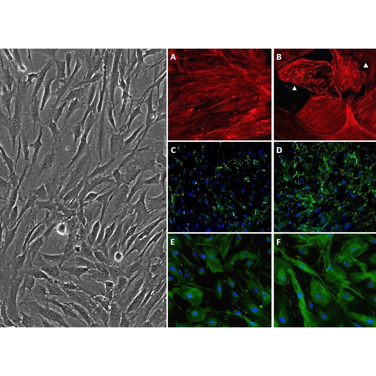
Human Trabecular Meshwork Cells: HTMC
Human Trabecular Meshwork Cells (HTMC) are specialized, multi-functional trabecular cells residing within the sponge-like connective tissue of the trabecular meshwork (TM), located in the anterior segment of the human eye.
Description
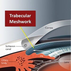
Human Trabecular Meshwork Cells (HTMC) are specialized, multi-functional trabecular cells residing within the sponge-like connective tissue of the trabecular meshwork (TM), located in the anterior segment of the human eye. The trabecular meshwork is organized into distinct structural zones—the uveal meshwork, the corneoscleral meshwork, and the juxtacanalicular tissue (JCT) — which form a series of extracellular matrix-coated beams and sheets. Positioned at the primary site of aqueous outflow resistance, each trabecular meshwork cell plays a critical role in maintaining intraocular pressure homeostasis.
These cells regulate the drainage of aqueous humor from the anterior chamber through the TM/JCT and across the inner wall endothelium into Schlemm’s canal. Rather than entering the bloodstream immediately, the fluid passes through Schlemm’s canal (collector channels/episcleral veins) endothelial cells — which form giant vacuoles and transcellular pores—and continues through collector channels into the episcleral veins to join the venous circulation. Outflow resistance is dynamically modulated by alterations in the contractility, focal adhesions, and cytoskeletal tension of the TM cells. Furthermore, because this region acts as a physical sieve, these cells exhibit a robust phagocytic nature that serves as a self-cleaning filter to remove cellular debris and maintain pathway patency, though this phagocytic capacity can vary significantly between TM cell subpopulations and declines with age and disease.
In the laboratory, establishing a primary cell culture using authentic human primary cells isolated directly from donor tissue provides an invaluable in vitro model for ophthalmic research. While some investigators use immortalized lines or animal tissue — such as harvesting enucleated mouse eyes to study the mouse eye or preparing a cell suspension from a mouse model — human primary cells remain the benchmark for capturing true human physiological thresholds.
When maintained in optimized trabecular meshwork cell medium, primary HTMC retain their sensitive responsiveness to mechanical stretch, steroid exposure, and growth factor signaling. This makes them indispensable reagents for modern drug development, enabling researchers to identify novel therapeutics that relax cell contraction, modify extracellular matrix (ECM) deposition, and safely lower intraocular pressure.
-
Pathogenesis of Glaucoma and TM Dysfunction: Progression of severe ocular disease is frequently rooted in a profound TM dysfunction. In glaucoma, a leading cause of irreversible blindness worldwide, the delicate homeostatic balance of the human eye is disrupted. A decrease in aqueous outflow facility leads to a pathological elevation of intraocular pressure, which places mechanical and ischemic stress on the posterior segment, resulting in the progressive loss of retinal ganglion cells. At the cellular level, this TM dysfunction is characterized by progressive trabecular meshwork cell loss and the accumulation of senescent cells, which compromises the tissue’s overall filtering capacity.
-
Extracellular Matrix Remodeling and Stiffening: During chronic glaucoma, HTMC undergo extensive phenotypic shifts. The cells exhibit pathological responses to elevated transforming growth factor-beta 2 (TGF-β2), which drives increased fibrosis, excessive fibronectin accumulation, and the upregulation of ECM cross-linking enzymes. This severe cytoskeletal reorganization causes the normally compliant trabecular beams to become rigid and stiff, permanently increasing fluid resistance before it reaches the inner wall tissue of Schlemm’s canal.
-
Mitochondrial Defects and Oxidative Stress: Sustained intraocular hypertension and environmental insults cause significant metabolic stress within TM cells. Glaucomatous tissues show pronounced mitochondrial defects, altered baseline signaling pathways, genomic DNA defects, and extensive oxidation damage. For instance, chronic hypoxia or oxidative stress increases localized DNA methylation, altering gene expression profiles and blunting the cells’ normal homeostatic repair mechanisms.
-
Pan-Tissue Heterogeneity and Endothelial Crosstalk: While frequently described as “endothelial-like,” TM cells are a heterogeneous population possessing myofibroblastic, macrophage-like, and endothelial-like features depending on their specific region and active activation state. Maintaining regular fluid clearance requires strict paracrine coordination between these heterogeneous trabecular cells and the inner wall endothelium of Schlemm’s canal. The endothelial cells lining the canal facilitate fluid transit into downstream venous channels, and investigators use co-culture arrays to study how growth factor secretion and mechanical signaling loops between HTMC and the canal’s inner wall govern global barrier permeability.
-
Regenerative Medicine and Advanced Nanoparticle Delivery: Given the finite lifespan and age-related decline of native trabecular cells, researchers are actively exploring stem-cell-based therapies to restore meshwork cellularity. Investigators evaluate how healthy stem cells can be differentiated into functional TM cells to repopulate damaged tissue. Concurrently, primary HTMC serve as a vital testing platform for optimizing non-viral gene delivery system designs, using targeted nanoparticles to alter gene expression, reduce contractility, and reverse fibrotic tissue remodeling directly at the drainage site.
Because TM cells are highly heterogeneous and can share baseline markers with adjacent tissues, primary trabecular meshwork cell populations maintained in specialized trabecular meshwork cell medium are typically validated using a combined panel of general, inducible, and functional assays rather than relying on a single isolated marker:
-
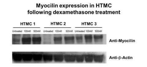
Dexamethasone Responsiveness & MYOC Upregulation: A classic hallmark used to confirm cell identity in vitro. Exposure to glucocorticoids induces the formation of cross-linked actin networks (CLANs) and upregulates the expression of Myocilin (MYOC). While MYOC mutations are known to cause specific hereditary forms of glaucoma, its acute upregulation in healthy cell culture serves as a key phenotypic identifier.
-
Phagocytic Activity Assays: Verified by documenting the cell’s baseline functional capacity to ingest fluorescently labeled microspheres or cellular debris over a timed assay.
-
Extracellular Matrix and Activation Markers: Validated through the expression of matrix Gla protein (MGP), aquaporin 1 (AQP1), chitinase-3-like-1 (CHI3L1), and alpha-smooth muscle actin (α-SMA) to evaluate the cell’s baseline activation or myofibroblastic state.
-
Endothelial Marker Context: Endothelial markers, such as VE-cadherin, must be interpreted with caution; while they are sometimes detected under specific culture conditions due to the tissue’s heterogeneous nature, they are primarily shared with or unique to the neighboring inner wall endothelium of Schlemm’s canal.
Human Trabecular Meshwork Cells (HTMC) are endothelial-like cells in a sponge-like connective tissue located near the front of the eye. HTMC make layers of beams, part of a fibrous basement membrane containing extracellular matrix and cells. In this area of high outflow resistance, HTMC regulate eye pressure by controlling drainage of fluid into tubes that flow into the bloodstream. Outflow is mediated by alterations in contractility and tension of HTMC, which also serve as a self-cleaning filter due to their phagocytic nature. Live cell imaging of the cytoskeleton provides valuable information on actin dynamics in HTMC. Other research aims to identify treatments that relax HTMC contraction to increase fluid outflow and lower eye pressure. Specific targeting of HTMC could also play a clinical role by increasing therapeutic efficacy of nanoparticles for gene delivery.
Damage and dysfunction of HTMC have clinical significance. For instance, hypoxia increases DNA methylation, accompanied by altered gene expression. During the normal aging process, HTMC number decreases, and senescent cells accumulate. In glaucoma, a leading cause of irreversible blindness, decreased fluid outflow causes an elevation of intraocular pressure and progressive loss of retinal ganglion cells. Physical changes to the TM include increased fibrosis, fibronectin accumulation, and expression of ECM cross-linking enzymes. This cytoskeletal reorganization and cell loss causes the TM to become rigid and stiff. Other glaucoma-related dysfunctions include mitochondrial defects, altered signaling pathways, elevated TGF-β2, genomic DNA defects, and oxidation damage.
Myocilin expression in Trabecular Meshwork Cells was analyzed following dexamathone treatment. After cells reach 70-80% confluency, HTMC cells were treated with 100nM and 500nM of dexamethasone for 6 days in DMEM with 1% FBS. Cell lysates were analyzed for Myocilin and beta-Actin expression by Western Blot.
Details
| Tissue | Normal healthy human adult eyes | |
|---|---|---|
| QC | No bacteria, yeast, fungi, mycoplasma, virus | |
| Character | Positive for Fibronectin | |
| Bioassay | Attach, spread, proliferate in Growth Med | |
| Cryovial | ~500,000 HTMC (1st passage) in Freezing Medium w/ 10% FBS, 10% DMSO | |
| Kit | Cryovial frozen HTMC, Growth Medium (631-500), Subculture Rgnt Kit (090K) | |
| Proliferating | Shipped in HTMC Growth Medium, psg 2, flasks or plates | |
| Doublings | At least 12 | |
| Applications | Laboratory research use only (RUO). Not for human, clinical, diagnostic or veterinary use. |
Resources
FAQs
Need More Help?
Visit our comprehensive FAQ page for detailed answers to common questions.
Need More Help?
Visit our comprehensive FAQ page for detailed answers to common questions.
Primary Cell FAQs Powered by Bioz