Canine Corneal Keratocytes: CnCK
Description
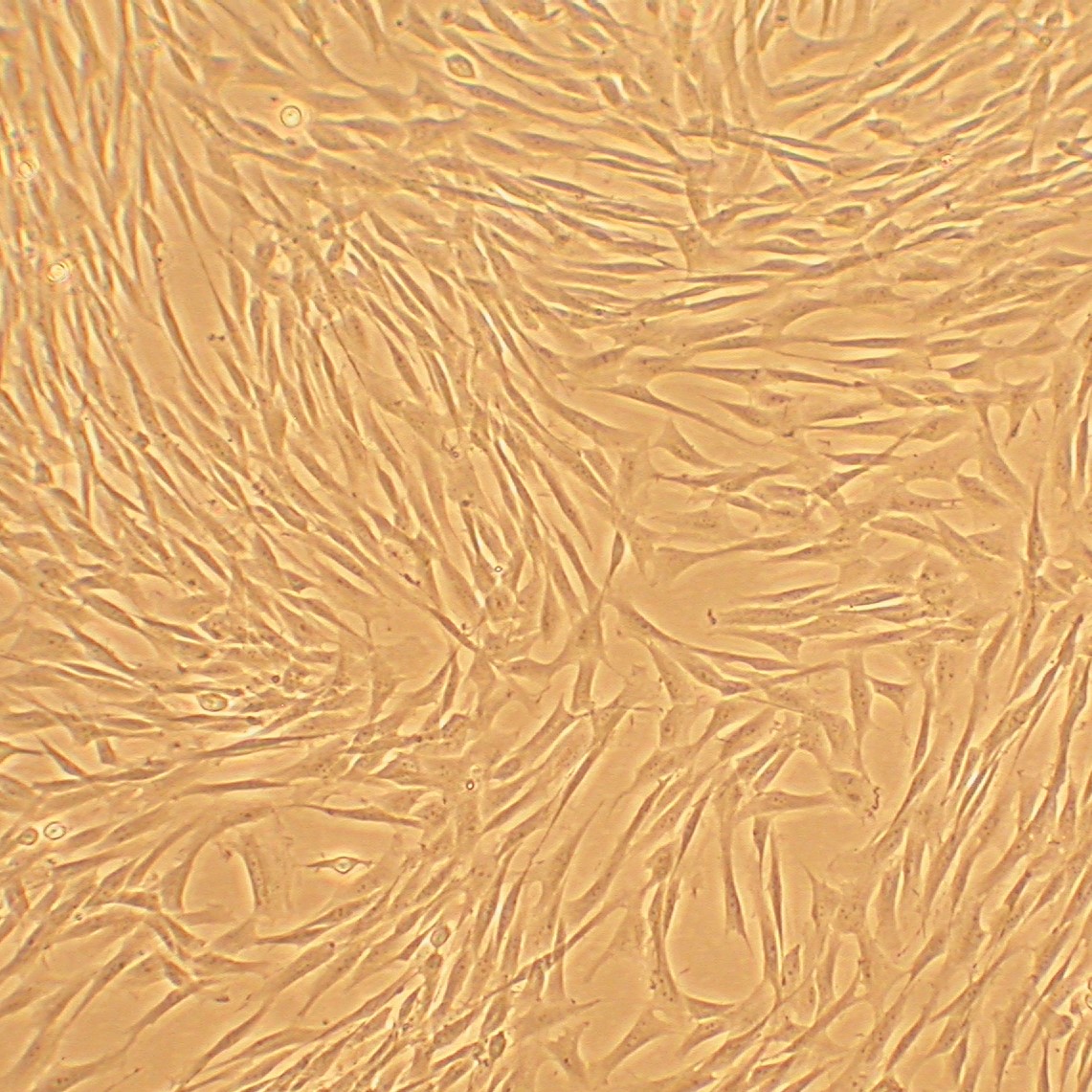
Canine Corneal Keratocytes (CnCK) are primary cells derived from canine eye. Corneal Keratocytes are neural-crest derived mesenchymal cells that populate the corneal stroma. The stroma is comprised of a type I and type V collagen fibers arranged as lamellae, makes up majority of the corneal thickness responsible for promoting corneal transparency. Upon corneal injury, keratocytes are stimulated to either undergo apoptosis or transition into repair pheanotypes to promote corneal regeneration.
Canine Corneal Keratocytes (CnCK) cultured in Corneal Keratocyte Growth Medium are able to propagate to 10 population doublings when cultured under the recommended conditions. CnCK could be used as an important in vitro cell model to study keratocyte differentiation, corneal injury and related diseases.
Details
| Tissue | Normal healthy canine cornea | |
|---|---|---|
| QC | No bacteria, yeast, fungi, mycoplasma, virus | |
| Bioassay | Attach, spread, proliferate in Growth Med | |
| Cryovial | 500,000 CnCK (1st passage) frozen in Basal Medium w/ 10% FBS, 10% DMSO | |
| Kit | Cryovial frozen CnCK (Cn632-05), Growth Medium (Cn6111-500), Subculture Rgnt Kit (090K) | |
| Proliferating | Shipped in Tsfr Med, psg 1, flasks or plates | |
| Doublings | At least 15 | |
| Applications | Laboratory research use only (RUO). Not for human, clinical, diagnostic or veterinary use. |
Resources
FAQs
Need More Help?
Visit our comprehensive FAQ page for detailed answers to common questions.
Need More Help?
Visit our comprehensive FAQ page for detailed answers to common questions.
Primary Cell FAQs Powered by Bioz