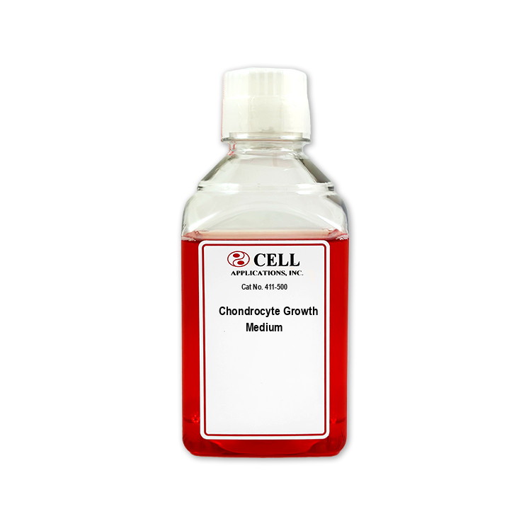
Human Chondrocyte Media for use with cartilage-producing cells.
Human Chondrocyte Media
HC Growth Medium: All-in-one ready-to-use
Type: Growth Medium
Size: 500 ml
Price: $147.00
Description

Human Chondrocyte Media is a specialized culture media designed to support chondrocytes, including articular chondrocytes derived from specialized cartilage in the diathrodial joint. In the body, chondrocytes reside within cartilage and maintain its load-bearing extracellular environment by regulating extracellular matrix production and turnover. Key matrix macromolecules characteristic of healthy hyaline cartilaginous tissue include type II collagen and aggrecan, which together help endow cartilage with compressive resilience and resistance to deformation. Because cartilage is avascular, nutrient and waste exchange rely heavily on diffusion, so cultured chondrocytes are sensitive to changes in the culture environment; maintaining appropriate growth conditions supports cellular viability while preserving key phenotypic traits of normal cartilage.
In research, Human Chondrocyte Media is used with primary chondrocyte sources, including primary chondrocyte and chondrocyte implantation workflows, as well as experimental systems that examine cartilage repair and remodeling processes in joint tissues. Chondrocytes can show phenotype shifts, including transition toward activated chondrocytes and altered matrix output as culture conditions vary; targeted cell culture conditions help support sustained performance of primary cells and improve consistency across experiments involving cell models. The media also supports studies of senescence and injury- or inflammation-associated changes in chondrocyte behavior, which is central to modeling disease progression in joint biology and joint mechanobiology.
Human Chondrocyte Media contributes to these research applications by providing an optimized culture media environment and appropriate media supplement composition to sustain expected chondrocyte growth and matrix-related functions during cell culture and 3d cell culture approaches. It supports experimental endpoints that depend on matrix biosynthesis and cartilage-like phenotypes, such as cartilage repair assays and evaluations of extracellular matrix production by monitoring matrix macromolecules output, including type II collagen and aggrecan expression. In addition, the media is commonly used in translationally relevant studies that incorporate mesenchymal stem cell–driven chondrogenic differentiation toward chondrocyte lineages, enabling comparisons between differentiated cells and mature chondrocytes within joint tissue models.
Finally, the same focus on supporting consistent cartilage-producing cells makes the medium useful for engineered and differentiation workflows, including studies using cell engineering tools and phenotypically stable cartilage-forming systems. In engineered platforms and differentiation experiments, reliable culture conditions help maintain chondrocyte performance and phenotype during processes that include stem cell–to–chondrocyte lineage transitions, and may be integrated with explants and other tissue-derived starting materials to more faithfully model specialized cartilage architecture and function in vitro, including cell biology experiments designed to quantify matrix remodeling, degradation, and regeneration in the joint environment.
Specialized media, with ingredients and supplements optimized for the unique requirements of Human Chondrocytes (HC), providing optimal culture conditions, cell health, data integrity and reproducibility.
Use with:
- Human Chondrocytes – Osteoarthritis: HC-OA
- Human Chondrocytes – Re-Differentiated: RD-HC
- Human Chondrocytes – Rheumatoid Arthritis: HC-RA
- Human Chondrocytes: HC
CAI media are tested for sterility in order to confirm no bacteria, yeast or fungi contaminate the solutions. The products undergo further quality control for correct pH, osmolality and lack of endotoxins. A panel of different bioassays affirm the media sustain a proper environment for expected cell-type-specific culture, growth, plating, karyotype, physiology, morphology, viability, population doublings, surface markers, cryopreservation, differentiation and/or induction.
Laboratory research use only (RUO). Not for human, clinical, diagnostic or veterinary use.
Details
Chondrogenesis Kit (Cat. No. 072K)
| Kit Component | Cat. No. | Storage | |||
|---|---|---|---|---|---|
| Sodium Alginate Solution | 072-25 | 25C | |||
| Sodium Chloride Solution | 074-100 | 25C | |||
| Calcium Chloride Solution | 071-100 | 25C | |||
| Depolymerization Solution | 073-50 | 25C | |||
| Chondrocyte Differentiation Medium | 411D-250 | 4C |
Sodium Alginate Solution
Cat. No. 072-25 / 25 ml
Sterile 1.2% solution, ready-to-use
Sodium Chloride Solution: NaCl
Cat. No. 074-100 / 100 ml
Sterile filtered 0.155 M saline solution, ready-to-use
Calcium Chloride Solution: CaCl2
Cat. No. 071-100 / 100 ml
Sterile, ready-to-use
Depolymerization Solution: DS
Cat. No. 073-50 / 50 ml
Sterile-filtered sodium citrate solution, ready-to-use
Chondrocyte Differentiation Medium
Cat. No. 411D-250
Ready-to-use
Chondrocytes cultured in monolayer quickly lose their phenotypic characteristics and no longer express Collagen type II and sulfated proteoglycan. Encapsulation of de-differentiated chondrocytes for 3 to 4 weeks in alginate beads will induce re-differentiation and ECM production in chondrocytes.
- Calcium Chloride Solution is formulated to induce polymerization of Sodium Alginate.
- Chondrogenesis Kit contains all media & solutions for the encapsulization of chondrocytes in alginate beads
- Sodium Alginate encapsulates chondrocytes to maintain their phenotypic characterization after isolation or to induce cultured chondrocytes to re-differentiate to chondrocytes.
- Depolymerization Solution (DS) consists of sodium citrate, and dissolves alginate beads to release re-differentiated chondrocytes.
Instructions for Releasing Re-differentiated Chondrocytes from Alginate Beads
- Remove Chondrocyte Differentiation Medium
- Wash the beads 2 times with 0.155 M NaCl
- Add 3 volume of Depolymerizatiojn Solution per volume of packed alginate beads
- Mix at room temperature for 30 minutes
- Apply gentle agitation until the beads have completely dissolved
- Pellet re-differentiated chondrocytes at 1000 rpm for 10 minutes
- Wash the cells 2 times with 0.155 M NaCl
 Powered by Bioz