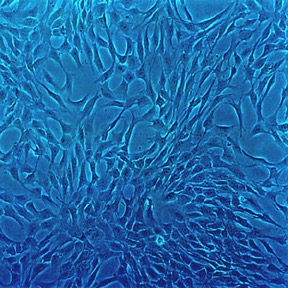
Human Chondrocytes: HC
Human Chondrocytes (HC) are found within healthy articular cartilage.
Description
Human Chondrocytes (HC) — also referred to as human articular chondrocytes or simply as a specialized cartilage cell type — are the sole resident cell population found within healthy articular cartilage. Unlike a transformed cell line, these primary chondrocytes retain their native genetic architecture and pathobiological memory in vitro. Structurally characterized by a spherical morphology in vivo, these isolated chondrocytes are the exclusive engine behind the biosynthesis, structural turnover, and homeostatic maintenance of the dense extracellular matrix. To preserve the specialized chondrocyte phenotype and minimize early dedifferentiation into an elongated, fibroblast-like morphology, investigators rely on a highly optimized chondrocyte medium. This specialized liquid environment supports stable gene expression and maintains a high percentage of viable cells, helping to extend their finite lifespan in culture.
In the human body, these articular chondrocytes are embedded within the unvascularized, uninnervated hyaline cartilaginous tissue that caps the articulating ends of bones in diarthrodial joints. Because this tissue lacks a direct blood supply, the cells reside in a low-oxygen environment and rely entirely on diffusion through the matrix to receive nutrients and clear metabolic waste. To evaluate structural joint health, investigators map how HC interact with neighboring cell types, including the subchondral bone matrix and the synovial lining layer. In commercial and academic research, a cell suspension of primary HC is typically characterized by checking the total cell count and ensuring the cells express structural markers that distinguish them from generalized mesenchymal stem cells or sublining stromal cells.
The primary biological function of healthy HC is to maintain the structural integrity of the joint by regulating a precise balance of matrix macromolecules. They synthesize and organize a complex structural network dominated by a specific collagen type (primarily Type II collagen), aggrecan, and highly sulfated glycosaminoglycans that give the tissue its compressive resilience. However, during mechanical overload or chronic joint inflammation, their function undergoes a pathological shift. Activated chondrocytes downregulate anabolic matrix synthesis and transition into a destructive catabolic state. Driven by interconnected inflammatory and signaling networks, they upregulate a robust panel of matrix metalloproteinases (such as MMP-3 and MMP-13) and aggrecanases (ADAMTS4/5) that systematically drive cartilage degradation and joint destruction.
In laboratory settings, a primary chondrocyte culture serves as a translationally rigorous human model for studying joint mechanobiology, mapping arthropathy progression, and validating advanced pipelines in tissue eng and regenerative medicine. Investigators must note that optimized primary media slow down but do not fully prevent replicative senescence; primary chondrocytes possess a limited proliferative capacity, and their progression toward senescence is heavily influenced by donor age, passage number, and specific culture conditions.
Researchers use these cells as a gold-standard biological benchmark to evaluate the efficiency of directing pluripotent or mesenchymal stem cells along the path of chondrogenic differentiation. Furthermore, because they produce authentic engineered cartilage, primary HC are critical for optimizing protocols like autologous chondrocyte implantation to repair a focal cartilage defect. By monitoring how variations in scaffold architecture or biochemical cues influence cell proliferation and chondrocyte maturation, orthopedic bioengineers can design biocompatible medical implants that successfully guide cartilage repair before irreversible structural failure occurs.
- Phenotypic characterization and transdifferentiation toward osteogenic/osteoblastic or hypertrophic chondrocyte-to-osteoblast-like phenotypes (endochondral ossification or hypertrophy).
- Others employ chondrocytes to describe the molecular biology of cell receptors, signaling cascades, cytokine activation and gene regulation.
- The cells are implicated in apoptosis, cytotoxicity, and cartilage degradation seen in joint disorders—including autoimmune conditions like rheumatoid arthritis, infectious models like Lyme disease-associated arthritis, and primarily degenerative conditions like osteoarthritis, where localized inflammatory processes contribute significantly to pathogenesis.
- By examining the effects of shear stress and mechanotransduction pathways, some hope to develop treatments to thwart erosive joint pathology.
- Some labs look at monoclonal antibody treatment, or inhibition of erosive matrix metalloproteinase enzymes, directed toward arthritis treatment.
- The cells also receive attention for potential clinical applications, since they adhere to medical implants and infiltrate scaffolds for cartilage regeneration.
Details
| Tissue | Normal healthy human articular cartilage | |
|---|---|---|
| QC | No bacteria, yeast, fungi, mycoplasma, virus | |
| Bioassay | Attach, spread, proliferate in Growth Med | |
| Cryovial | 500,000 HC (1st passage) frozen in Basal Medium w/ 10% FBS, 10% DMSO | |
| Kit | Cryovial frozen HC (402-05), Growth Medium (411-500), Subculture Rgnt Kit (090K) | |
| Proliferating | Shipped in Gr Med, 2nd psg (flasks or plates) | |
| Doublings | At least 10 | |
| Applications | Laboratory research use only (RUO). Not for human, clinical, diagnostic or veterinary use. |
Resources
FAQs
Need More Help?
Visit our comprehensive FAQ page for detailed answers to common questions.
Need More Help?
Visit our comprehensive FAQ page for detailed answers to common questions.
Primary Cell FAQs Powered by Bioz