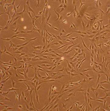
Human Chondrocytes: Osteoarthritis: HC-OA
Human Chondrocytes: Osteoarthritis (HC-OA) are specialized cells isolated directly from the diseased cartilage tissue of donors diagnosed with osteoarthritis
Description
Human Chondrocytes: Osteoarthritis (HC-OA) — also known as osteoarthritis articular chondrocytes or primary OA chondrocytes — are specialized resident cells isolated directly from the diseased cartilage tissue of donors diagnosed with osteoarthritis. Unlike a continuous cell line, these primary chondrocytes retain their native pathobiological memory, epigenetic modifications, and disease-associated metabolic features ex vivo. In a healthy joint, mature chondrocytes are responsible for maintaining the homeostatic structural network of hyaline cartilage. However, inside the joint of OA patients, the cells undergo a profound phenotypic shift. To study these disease mechanics in vitro without inducing compounding artifactual changes, researchers utilize a specialized chondrocyte medium. This system allows them to stabilize the cells for downstream molecular biology assays and track how the chondrocyte phenotype responds to targeted therapeutic interventions.
In the human body, these cells reside within the load-bearing, unvascularized OA cartilage caps that line the articulating surfaces of arthritic joints. In this altered microenvironment, the cells are exposed to a complex combination of mechanical overload and localized biochemical stress. While healthy human articular chondrocytes are shielded from systemic circulation, the progression of OA permits an influx of inflammatory mediators secreted by neighboring tissues, such as the hyperplastic synovial membrane and infiltrating immune cells. This creates a highly catabolic milieu where the isolated cells are constantly exposed to pro-inflammatory cytokines like TNF- and IL-1β, which accelerates cartilage degradation.
The primary biological function of articular chondrocytes is to balance the synthesis and breakdown of the extracellular matrix, which is natively rich in type II collagen (specifically collagen type II) and aggrecan. In HC-OA, this homeostatic balance is fundamentally disrupted, causing catabolic matrix degradation to outpace anabolic matrix synthesis. Under chronic inflammatory stress, the cells undergo chondrocyte hypertrophy and transition into a state resembling a hypertrophic chondrocyte. Driven by interconnected inflammatory and signaling networks, these cells upregulate destructive matrix proteases. Specifically, aggrecanases (ADAMTS4/5) cleave aggrecan core proteins early in the disease, while interstitial collagenases—predominantly MMP-13—execute highly specific cleavages within the structural triple-helix of type II collagen fibers. Furthermore, a substantial fraction of the population shifts into senescent cells or undergoes chondrocyte apoptosis, which facilitates subchondral bone remodeling, matrix calcification, and the permanent loss of normal joint mechanics.
In laboratory settings, a primary culture of HC-OA serves as a translationally rigorous human model for interrogating degenerative pathways, mapping mechanotransduction loops, and validating disease-modifying osteoarthritis drugs (DMOADs). Researchers utilize these cells to evaluate advanced cartilage repair strategies and to understand the mechanisms of cartilage formation. For instance, investigators deploy HC-OA in tissue engineering (tissue eng) pipelines to test whether specialized biomimetic scaffolds or nanoparticles can suppress terminal chondrocyte hypertrophy and restore a favorable anabolic balance.
Additionally, because these cells represent a definitive diseased state, they serve as an essential biological benchmark in regenerative medicine. Investigators must note, however, that because HC-OA function as a diseased benchmark, their responsiveness to regenerative cues, biochemical factors, and mechanical scaffolding may differ significantly from healthy chondrocytes. They are widely used alongside a donor mesenchymal stem cell population or healthy chondrogenic cells to evaluate how disease signaling alters cartilage regeneration, helping researchers design advanced therapies to treat a focal cartilage defect before irreversible structural failure occurs.
Details
| Tissue | Human articular cartilage from donor w/ osteoarthritis | |
|---|---|---|
| QC | No bacteria, yeast, fungi, mycoplasma, virus | |
| Bioassay | Attach, spread, proliferate in Growth Med | |
| Cryovial | 500,000 HC-OA (1st passage) frozen in Basal Medium w/10% FBS, 10% DMSO | |
| Kit | Cryovial frozen HC-OA (402OA-05), Growth Medium (411-500), Subcltr Rgnt Kit (090K) | |
| Proliferating | Shipped in Gr Med, 2nd psg (flasks or plates) | |
| Doublings | At least 10 | |
| Applications | Laboratory research use only (RUO). Not for human, clinical, diagnostic or veterinary use. |
Resources
FAQs
Need More Help?
Visit our comprehensive FAQ page for detailed answers to common questions.
Need More Help?
Visit our comprehensive FAQ page for detailed answers to common questions.
Primary Cell FAQs Powered by Bioz