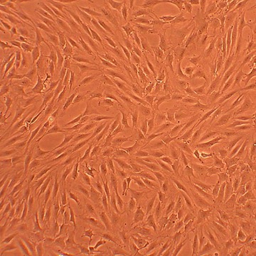
Human Chondrocytes-Rheumatoid Arthritis: HC-RA. Isolated from a donor patient with joint disease.
Human Chondrocytes: Rheumatoid Arthritis: HC-RA
Human Chondrocytes isolated from tissues affected by Rheumatoid Arthritis (HC-RA) are primary cells obtained from RA-affected cartilage.
Description
Human Chondrocytes isolated from tissues affected by Rheumatoid Arthritis (HC-RA) — also designated as rheumatoid arthritis articular chondrocytes — are primary cells obtained from RA-affected cartilage. Rheumatoid arthritis is a systemic, chronic autoimmune disease characterized by the generation of autoantibodies and the activation of autoreactive T cell subsets, which culminate in the hyperproliferation of synovial fibroblasts (pannus formation) and immune cell infiltration. While healthy chondrocytes maintain an anabolic-catabolic equilibrium within the cartilage matrix, the microenvironment in RA pathogenesis is drastically reshaped by an intense proinflammatory cytokines storm. Consequently, primary HC-RA serve as a highly relevant human in vitro model for studying the biological effects of chronic inflammation on cell function, evaluating metabolic differences between healthy and pathological joints, and validating targeted therapies for patients. These cells are typically maintained in a specialized chondrocyte medium to preserve their phenotype for research.
In the rheumatoid joint, HC-RA are subjected to aggressive extrinsic pressures that override normal maintenance and drive cartilage degradation.
- The Pannus-Cartilage Interface: The hyperplastic synovial pannus directly invades the adjacent cartilage, exposing HC-RA to high concentrations of macrophage- and fibroblast-derived inflammatory cytokines found in the synovial fluid. These signaling molecules, which can originate from various cells including B cell and Th17 cells, act to amplify inflammatory responses. The local T cell activation and presence of Treg cells within this environment further modulate the severity of the joint damage.
- Matrix Breakdown: In response to these chronic exposures, the gene expression profiles within HC-RA are severely remodeled. The cells exhibit markedly increased expression and secretion of matrix-degrading enzymes—particularly interstitial collagenases and aggrecanases. This enzymatic activity acts in tandem with the invading pannus to accelerate the destruction of the cartilage matrix, characterized by the degradation of Type II collagen (or II collagen), ultimately leading to significant cartilage destruction and structural cartilage defect formation.
To fully map the complex dynamics of autoimmune joint destruction, investigators routinely deploy HC-RA within cross-disease comparative platforms.
- Disease-Specific Mechanisms: HC-RA are frequently utilized in parallel with healthy human chondrocytes and osteoarthritic chondrocytes. These comparative assays help researchers distinguish the pathways driven by the systemic, high-grade inflammatory environment of RA from the mechanically driven processes of osteoarthritis in the knee.
- Chondroprotective Validation: HC-RA serve as an essential discovery platform for evaluating localized, chondroprotective interventions. Researchers utilize these models to screen inhibitors, monoclonal antibodies, and advanced biomaterials aimed at preserving chondrocyte viability, preventing chondrocyte apoptosis, and restoring an anabolic balance. This research is crucial for identifying treatments that might one day improve outcomes in procedures like autologous chondrocyte implantation. By employing human-specific antibodies to quantify the expression of enzymes like MMP-1 or MMP-13, researchers can accurately track the progression of matrix breakdown and validate the efficacy of these novel therapeutics.
Human Chondrocytes Rheumatoid Arthritis (HC-RA) are derived from human articular cartilage of donors with rheumatoid arthritis (RA). Rheumatoid arthritis is an inflammatory disease that is characterized by overproliferation of synovial cells and their infiltration into adjacent joint tissues, accompanied by gradual destruction of cartilage and bone erosion. Normally, chondrocytes are responsible for producing and maintaining the extracellular matrix of cartilage, but in RA cartilage, the environment is changed significantly due to inflammation and the invasion of other cells. Therefore, HC-RA can be a useful model to study the effects of those changes on chondrocyte biology. In parallel with normal HC, HC-RA can be used to investigate the differences between normal and pathological joints and evaluate potential treatment strategies.
Characterization: Positive for aggrecan after differentiation
Details
| Tissue | Human articular cartilage, from donor w/ rheumatoid arthrits | |
|---|---|---|
| QC | No bacteria, yeast, fungi, mycoplasma, virus | |
| Bioassay | Attach, spread, proliferate in Growth Med | |
| Cryovial | 500,000 HC-RA (1st passage) frozen in Basal Medium w/10% FBS, 10% DMSO | |
| Kit | Cryovial frozen HC-RA (402RA-05), Growth Medium (411-500), Subcltr Rgnt Kit (090K) | |
| Proliferating | Shipped in Gr Med, 2nd psg (flasks or plates) | |
| Doublings | At least 10 | |
| Applications | Laboratory research use only (RUO). Not for human, clinical, diagnostic or veterinary use. |
Resources
FAQs
Need More Help?
Visit our comprehensive FAQ page for detailed answers to common questions.
Need More Help?
Visit our comprehensive FAQ page for detailed answers to common questions.
Primary Cell FAQs