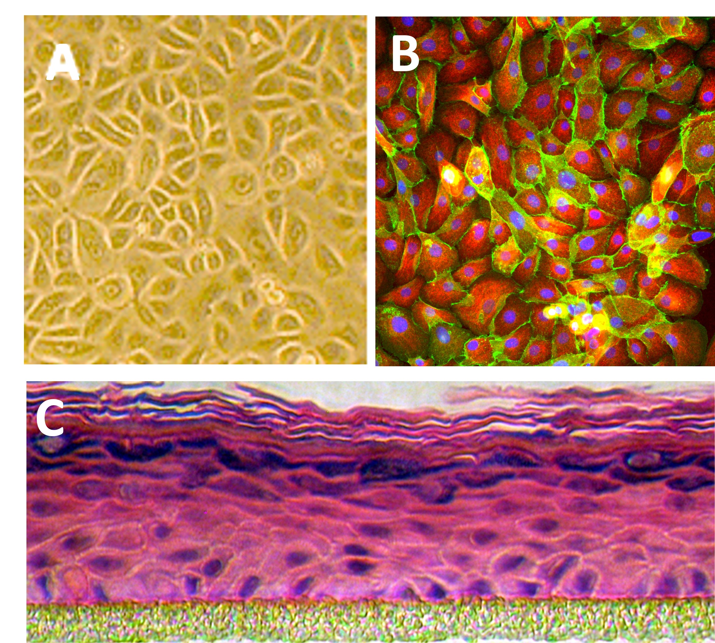
Human Epidermal Keratinocytes: HEK
Human Epidermal Keratinocytes (HEK) are keratinocyte cells from the epidermis, the outermost layer of the skin
Description
Human Epidermal Keratinocytes (HEK) — frequently studied as normal human epidermal keratinocytes or primary epidermal keratinocytes—comprise approximately 90% of all cells in the epidermis, the outermost layer of the skin. As a highly dynamic cell type, they transition from proliferative basal cells anchored to the basement membrane up through stratified layers to form the stratum corneum. This complex process of cellular differentiation constructs the body’s primary mechanical and permeability barrier, driven by the structured assembly of keratin intermediate filament proteins (keratins). In vivo, these human epidermal keratinocytes work in close tandem with neighboring melanocytes, absorbing melanin granules and positioning them as a protective nuclear shield against damaging ultraviolet (UV) radiation.
In the laboratory, utilizing human primary cells isolated directly from tissue offers distinct advantages for modeling human skin over lines like HaCaT, which is a spontaneously immortalized, non-transformed line that can exhibit altered baseline signaling profiles. Maintaining these primary cultures requires specialized media formulations, such as optimized Human EpiVita Media, to enhance their in vitro lifespan and preserve correct physiological responses. By choosing high-quality reagents and seeding them into appropriate culture vessels, researchers can investigate the delicate balance between keratinocyte proliferation and sequential differentiation. This establishes a high-fidelity platform to discover novel therapeutic targets, evaluate topical formulations, and map complex epidermal tissue dynamics.
-
Mechanisms of Keratinocyte Differentiation and Barrier Function: A core focus of keratinocyte culture is mapping the precise genetic cascades governing keratinocyte differentiation. When primary basal keratinocytes are grown on porous inserts and exposed to an air-liquid interface (ALI), they shift from active proliferation to a highly organized stratification program. This process closely mimics the in vivo basal layer maturation, driving the chronological expression of differentiation markers such as involucrin, loricrin, and filaggrin to form a functional, three-dimensional epidermal tissue model.
-
Wound Repair, Tissue Regeneration, and Chronic Diseases: Following physical trauma or barrier disruption, human epidermal keratinocytes rapidly alter their phenotype to initiate wound repair. They downregulate rigid cell-matrix attachments, migrate across the injured provisional matrix, and proliferate to re-epithelialize the tissue. Primary models allow investigators to dissect these migratory pathways and study chronic epidermal conditions like eczema or psoriasis, where aberrant cytokine signaling disrupts normal homeostasis and impairs barrier resolution.
-
Viral Infection, Pathogen Interactions, and Host Defense: As the body’s frontline shield, the epidermis is a primary site for viral infection and microbial colonization. Researchers utilize human epidermal keratinocytes to study the entry, replication, and pathogenesis of viruses like the Human Papillomavirus (HPV), which infects basal keratinocytes typically via microabrasions. Its lifecycle is linked to keratinocyte differentiation. Furthermore, these models are deployed to characterize how the epithelium secretes antimicrobial peptides in response to pathogenic bacteria, opportunistic yeast, or structural fungi.
-
Oncogenesis, Skin Cancer, and Genetic Target Validation: Primary epidermal models provide a crucial benchmark for studying genetic instability and the initiation of non-melanoma skin cancer, such as basal cell carcinoma and squamous cell carcinoma. By utilizing an advanced gene delivery system (such as lentiviral or CRISPR-based vectors), investigators can precisely alter target gene expression within primary cells. This allows them to analyze how specific oncogenic mutations bypass normal cell-cycle checkpoints and disrupt the balance between self-renewal and terminal exhaustion.
Details
| Tissue | Normal healthy human foreskin or adult skin | |
|---|---|---|
| QC | No bacteria, yeast, fungi, mycoplasma, virus | |
| Bioassay | Attach, spread, proliferate in Growth Med | |
| Cryovial | 500,000 HEK (Primary culture) frozen in Basal Medium w/ 10% FBS, 10% DMSO | |
| Kit | Cryovial HEK (102-05), Epi-Vita Growth Medium (141-500a for adult; 141-500 for fetal/neonatal), Subcltr Rgnt Kit (090K) | |
| Proliferating | In Serum-Fr Gr Med, psg 1, flasks or plates | |
| Doublings | At least 16 in keratinocyte media | |
| Applications | Laboratory research use only (RUO). Not for human, clinical, diagnostic or veterinary use. |
Resources
FAQs
Need More Help?
Visit our comprehensive FAQ page for detailed answers to common questions.
Need More Help?
Visit our comprehensive FAQ page for detailed answers to common questions.
Primary Cell FAQs Powered by Bioz