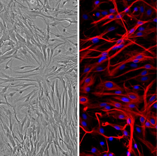
Rat Astrocytes: RA
Rat Astrocytes (RA) are highly specialized macroglial cells typically isolated from the cerebral cortex of rat brain tissue
Description
Rat Astrocytes (RA) are highly specialized macroglial cells typically isolated from the cerebral cortex of rat brain tissue, frequently sourced from specific strains such as Sprague Dawley. In the mammalian brain, astrocytes represent a major cell type among glial cells, with their relative abundance compared to partner neurons varying across specific brain regions and species. Characterized by their complex, highly branched, and star-shaped (stellate) morphology in vivo, these cells serve as indispensable architectural and functional anchors for neural tissue.
Astrocytes are known to operate as dynamic metabolic engines and active signaling hubs. In an astrocyte culture, these primary cells can retain astrocyte identity and transporter/expression profiles under optimized culture conditions, distinguishing them from immortalized cell lines or transformed glioblastoma models often used in cancer research. To maintain their non-reactive phenotype in vitro, they are typically maintained using a dedicated astrocyte medium animal system, specifically utilizing rat astrocyte growth medium, or rat astrocyte basal medium supplemented with a defined rat astrocyte growth supplement. Standard quality control protocols evaluate these primary cell products to ensure they are free of mycoplasma and other microbial contaminants, making them reliable tools intended for research use only and not for in vitro diagnostic procedures.
In the living organism, Rat Astrocytes (RA) are distributed throughout the central nervous system, extending elaborate processes that envelop both blood vessels and synaptic junctions. They are positioned within the neurovascular unit, where brain microvascular endothelial cells, pericytes, astrocytes, and the basement membrane function coordinately to maintain CNS privilege. Astrocytes are implicated in blood-brain barrier (BBB) regulation, though BBB-like interactions require appropriate co-culture or system context, as monocultured cells lack the necessary vascular partners to form active astrocytic endfeet interfaces.
Because astrocytes exhibit diverse properties and structural specializations depending on their exact anatomical location — such as the classic fibrous astrocytes in white matter versus protoplasmic astrocytes in grey matter — findings from numerous studies highlight that their baseline transporter profiles, metabolic constraints, and receptor densities are highly region-specific.
The primary biological function of native astrocytes is to regulate local homeostasis, coordinate synaptic transmission, and support normal astrocyte functions that directly impact neuron survival and synaptic plasticity. In vivo or in multi-lineage systems, astrocytes participate as full functional partners at the tripartite synapse. They express functional receptors for neurotransmitters like glutamate and -aminobutyric acid (GABA), and they clear excess glutamate from the synaptic cleft via high-affinity transporters like GLT-1 (EAAT2) and GLAST (EAAT1) to shield neurons from excitotoxic neural damage.
Additionally, astrocytes act as metabolic mediators; they can absorb glucose from the microvasculature to synthesize lactate, which is hypothesized to serve as an energy substrate for neighboring neurons, though the magnitude and presence of this metabolic transfer depends strongly on the experimental setup and cell-type composition. Through the controlled trophic factor release of molecules like thrombospondins and neurotrophins, astrocytes are also understood to help guide neurite outgrowth, provide neuronal guidance, and modulate synaptic remodeling in appropriate cellular environments.
In cellular neurobiology, neurotoxicology, and discovery pharmacology, RA can serve as a translationally relevant model system to explore glial biology, model brain injury phenotypes, and investigate complex pathological processes. Under conditions simulating ischemic stroke—such as in vitro oxygen-glucose deprivation (OGD) models—investigators use RA cultures to assess how specific interventions modulate cellular survival, metabolic indicators, or the release of pro-inflammatory cytokines like IL-1β and TNF-. Researchers also leverage RA to study reactive astrogliosis pathways, evaluating how chemical stress or structural injury prompts a transition into a reactive state that can either beneficially scar-bound a lesion or impede tissue regeneration depending on the specific microenvironmental stimuli involved.
Rat Astrocytes can be integrated into bioengineering workflows and advanced 3D cell culture setups. Investigators often pair them with electrospun nanofibrous matrices, hydrogels, or multi-electrode arrays to evaluate how successfully astrocytic scaffolds can support neurite outgrowth, establish physical tracks for neuronal guidance, or facilitate neuron regeneration models. To support these multi-cellular and bioengineered experimental designs, specialized associated products — including coating matrices, optimization reagents, and specific analytical tools — are frequently evaluated alongside these primary cell populations.
Biosensors and Lab-on-a-Chip Devices: Microfluidic biophysicists integrate RA into specialized multi-cellular lab-on-a-chip configurations, multi-electrode arrays, and high-throughput biosensors. These scaled technologies allow investigators to analyze real-time astrocytic responses to mechanical fluid shear, localized neurotoxins, or targeted drug delivery profiles, bypassing the limitations of traditional static plastic cultures.
- Assess neuroprotective capabilities of bioenergy stabilizers in an in vitro model of stroke
- Investigate the effects of astrocytes on brain microvascular endothelial cells in relation to iron transport pathway
Details
| Tissue | Normal healthy Rat brain | |
|---|---|---|
| QC | No bacteria, yeast, fungi, mycoplasma | |
| Cryovial | 500,000 RA (2nd passage) frozen in Basal Medium w/10% FBS, 10% DMSO | |
| Character | Positive for GFAP | |
| Bioassay | Attach, spread, proliferate in Growth Medium | |
| Kit | Cryovial frozen RA (R882A-05n), Growth Med (R821-500), Subcltr Rgnt Kit (090K) | |
| Proliferating | Shipped in Gr Med, 3rd psg (flasks or plates) | |
| Doublings | At least 10 | |
| Applications | Laboratory research use only (RUO). Not for human, clinical, diagnostic or veterinary use. |
Resources
FAQs
Need More Help?
Visit our comprehensive FAQ page for detailed answers to common questions.
Need More Help?
Visit our comprehensive FAQ page for detailed answers to common questions.
Primary Cell FAQs Powered by Bioz