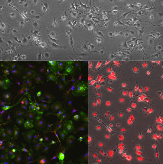
Rat Microglia Cells (RMcg) cultured 3 days (Top) and stained (Bottom) for CD11b/c (green) F4/80 (red), DAPI (blue) (L) and DiI-Ac-LDL (red) uptake (R).
Rat Microglia Cells: RMcg
Rat Microglia (RMcg) are specialized primary glial cells derived from the brain tissue of postnatal rats.
Description
Rat Microglia (RMcg) are specialized primary glial cells derived from the brain tissue of postnatal rats. As the resident brain macrophages, they represent a distinct cell type separate from macroglia (like astrocytes and oligodendrocytes). They are the primary immune cells of the central nervous system (CNS), forming an intricate neuro-glial cell network that maintains neural health.
While researchers frequently compare them to microglial cells in mouse models or aged mice, RMcg offer an invaluable, biologically precise mammalian model for in vitro experimentation. Under healthy conditions, these cells exhibit a highly branched, resting microglia morphology, allowing them to continuously monitor their microenvironment.
In vivo, microglia originate from yolk sac progenitors during early embryonic development and populate the brain parenchyma. They are distributed throughout virtually every brain region, including the cerebral cortex (where they are studied as cortical microglia), hippocampus, and spinal cord.
The CNS is strictly separated from systemic circulation and peripheral immune elements (like circulating T cells) by the blood-brain barrier (BBB). Because the BBB limits peripheral immune access, these glial cell types serve as the dominant force in brain immune surveillance. While other resident populations like border-associated macrophages exist, microglia are the principal sentinels within the immune-privileged space of the brain.
Microglia are highly dynamic cells with multifaceted roles spanning from early brain development to the maintenance of cognitive function in adulthood.
1. Surveillance and Homeostasis
In a baseline state, they constantly sample the environment to modulate neuronal activity and manage synaptic pruning. They respond dynamically to molecular signals, where changing expression levels of surface receptors regulate their baseline behavior.
2. Microglial Phagocytosis & Clearance
When tissue damage occurs, microglia trigger microglial phagocytosis to clear cellular debris, misfolded protein aggregates, and cells undergoing programmed cell death. This can occur via several pathways, including Fc-mediated phagocytosis, which relies on specific antibody-receptor engagement.
3. Activation and Multi-Dimensional Spectrum
Upon detecting injury, pathogens, or a disrupted BBB, microglia undergo microglial activation. This process alters their gene expression profiles and transforms their physical structure from a ramified shape to a motile, amoeboid form.
Historically, microglia polarization has been taught using a simplified, binary model borrowed from peripheral macrophages:
M1 Polarization (Classical Activation): A state where activated microglia release pro-inflammatory cytokines (such as IL-1β, TNF-α, and IL-6) and other cytokine signaling molecules to combat acute infection.
M2 Microglia (Alternative Activation): A state focused on neuroprotection and tissue repair, releasing growth factor molecules to resolve inflammation.
Modern Scientific Nuance: Modern neuroimmunology recognizes that this rigid binary classification is an oversimplification that does not capture the full complexity of in vivo behavior. Rather than discrete phenotypes, microglia activation represents a highly fluid continuum. The actual microglia phenotype exists on a multidimensional spectrum, with considerable heterogeneity depending on the specific disease context, tissue region, and unique local stimuli.
Because human CNS tissue is difficult to obtain, isolated Rat Microglia (RMcg) serve as a critical in vitro model to study neuroinflammation, neurodevelopment, and neurodegenerative diseases.
Phenotypic Characterization
Scientists rely on specific microglial marker profiles to confirm the purity of RMcg cultures. Tools like flow cytometry and immunocytochemistry are used to analyze the expression of target proteins (such as CD11b, CD68, and Iba1) to evaluate their activation state and assess the purity of the isolated cells against contaminating astrocytes or neurons.
Disease Modeling & Therapeutic Testing
RMcg are widely used to investigate how microglial responses shift during pathological aging, mimicking the altered, chronically inflamed states seen in aged microglia. Researchers manipulate these cultures to study how changes in genes and downstream signaling cascades impact survival and neurotoxicity. Furthermore, techniques involving microglia depletion in co-culture systems allow scientists to isolate variables, determining exactly how a lack of microglial support affects neighboring neurons and overall network stability.
Rat Microglia (RMcg) are derived from the brain of normal postnatal day 1 rats by standardized methods. When revived and plated and cultured under recommended conditions, RMcg form adherent culture.
Microglia, originated from the hematopoietic stem cells in bone marrow, are the resident macrophages in the central nervous system (CNS) which is separated from the rest of the body due to the presence of blood-brain barrier. Microglia actively survey the surrounding area and respond by scavenging damaged neural cells, plaques, and infectious agents. Due to their functions in immune response and maintaining homeostasis in the CNS, microglia have been implicated in neurodevelopment, CNS plasticity and repair, neuroinflammation, aging and neurodegeneration, neuropathic pain, and infections.
Characterization: RMcg stain positive for F4/80 and CD11b/c, and uptake DiI-Ac-LDL
Details
| Tissue | Normal, healthy Rat brain | |
|---|---|---|
| QC | No bacteria, yeast, fungi, mycoplasma | |
| Character | Positive for F4/80 and CD11b/c | |
| Bioassay | Plate, spread on CC ware & uptake DiI-Ac-LDL | |
| Cryovial | 1M RMcg frozen in Macrophage & Microglia Cell Freezing Med (043-50) | |
| Kit | Cryovial Frozen RMcg (R8816K-10n), Macrophage Cltr Med Kit (R619K-100), Coating Soln (1021-5) | |
| Doublings | N/A – Microglia Cells don’t proliferate in vitro | |
| Applications | Laboratory research use only (RUO). Not for human, clinical, diagnostic or veterinary use. |
Resources
FAQs
Need More Help?
Visit our comprehensive FAQ page for detailed answers to common questions.
Need More Help?
Visit our comprehensive FAQ page for detailed answers to common questions.
Primary Cell FAQs