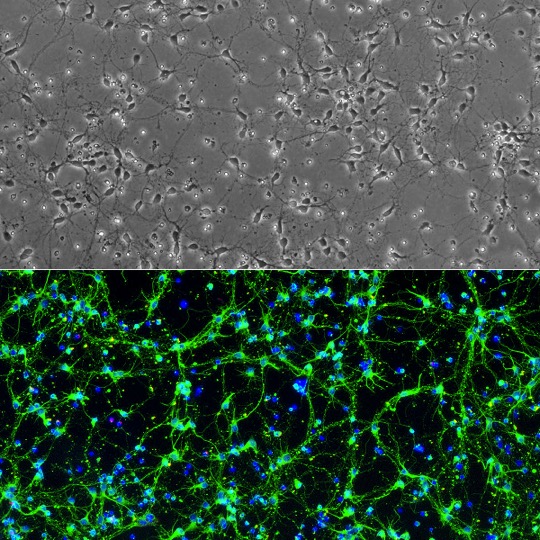
Cryopreserved Rat Midbrain Neurons (RMbN) revived and cultured for 6 days (top), stained (bottom) on day 7 for β III-Tubulin (green), GFAP (red), and DAPI (blue).
Rat Midbrain Neurons: RMbN
Rat Midbrain Neurons (RMbN) are primary neural cells derived from the midbrain region of the embryonic rat brain (typically gestational day 15).
Description
Rat Midbrain Neurons (RMbN) are primary neural cells derived from the embryonic rat brain (typically gestational day 15). When cultured under recommended conditions, these populations arborize and form complex neurite networks, staining positive for classical neuronal markers like class III beta-tubulin. The midbrain is a critical anatomical region within the mammalian brain that regulates motor control, sensory processing, and arousal systems, housing specialized structures such as the substantia nigra pars compacta and the ventral tegmental area.
Cultures derived from the midbrain — particularly the ventral mesencephalon — are often enriched for dopaminergic neurons and midbrain dopaminergic neurons, enabling studies of reward circuitry, motor control pathways, and neurodegenerative conditions like Parkinson’s disease. While in vitro primary cultures provide controlled models for cellular assays, researchers occasionally compare findings or complement primary systems with alternative platforms, such as stem cells, organoids, or comparative models derived from mice (and contrasted with circuits involving the cerebral cortex), to investigate broader aspects of brain development and neurodegenerative pathology.
Cultures of primary Rat Midbrain Neurons (RMbN) serve as a versatile experimental platform for evaluating functional actions, monitoring target mRNA levels, and evaluating the effects of neuroactive substances. These models can be applied across a wide range of analytical workflows — including toxicity testing, immunostaining, live-cell imaging, and electrophysiology — while maintaining stringent quality control to screen for common culture contaminants such as mycoplasma and preserve experimental fidelity.
Rat Midbrain Neurons (RMbN) are derived from midbrains of normal embryonic rat (gestation day 15) by standardized methods. When cultured under the recommended conditions, RMbN arborize and form complex neurite network in one week. RMbN Stain positive for β III-Tubulin.
The midbrain is a small part of the brain yet plays important functions in visual and auditory systems, motor control, alertness, sleep/wake, and temperature regulation. It contains the substantia nigra, a darkly pigmented region with high number of dopaminergic neurons, which is closely associated with motivation and habituation. Atrophies and diseases at midbrain seen in conditions such as Parkinson’s disease, familial amyotrophic lateral sclerosis, and progressive supranuclear palsy affect balancing, gait and muscle control. Studying midbrain neurons in those disease conditions will allow a better understanding of the disease mechanisms, physiopathologies, and advancement in drug development and therapies. Cultures of midbrain neurons can be applied for a variety of experiments including toxicity test, immunostaining, live cell imaging, co-culturing, electrophysiology, and more.
Details
| Tissue | Normal healthy rat midbrain | |
|---|---|---|
| QC | No bacteria, yeast, fungi, mycoplasma | |
| Character | Positive for β-III Tubulin | |
| Bioassay | Plate on Poly-D-Lysine coated surface, arborize to form neurite network in Culture Med | |
| Cryovial | 1M RMbN in Serum-Free Frzng Med (042-50) | |
| Kit | Cryovial frzn RMbN, Neuron Ctng Soln I (027-05), Plating (R817P-10) & Cultr Med (R817-100) | |
| Doublings | N/A Neurons don’t proliferate in vitro | |
| Applications | Laboratory research use only (RUO). Not for human, clinical, diagnostic or veterinary use. |
Resources
FAQs
Need More Help?
Visit our comprehensive FAQ page for detailed answers to common questions.
Need More Help?
Visit our comprehensive FAQ page for detailed answers to common questions.
Primary Cell FAQs