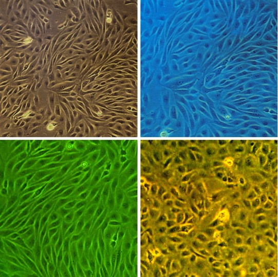
Bovine Brain Microvascular Endothelial Cells: BBMVEC. Isolated from small cerebral blood vessels.
Bovine Brain Microvascular Endothelial Cells: BBMVEC
Bovine Brain Microvascular Endothelial Cells (BBMVEC) are a specialized primary cell type isolated from the bovine brain microvessel network.
Description
Bovine Brain Microvascular Endothelial Cells (BBMVEC) are a specialized primary cell type isolated from the bovine brain microvessel network, providing an essential in vitro system for studying cerebrovascular biology and modeling the blood brain barrier (BBB). Unlike macrovascular or peripheral endothelial cells, these brain endothelial cells display BBB-like endothelial properties — such as low pinocytic activity and reduced paracellular transport — depending on culture conditions, alongside specialized transport functions relevant to the central nervous system.
When established as a confluent BMEC monolayer, these primary cultures can develop intercellular tight junctions (characterized by the localization of key tight junction protein markers such as occludin and ZO-1). This structural organization supports barrier-function assays (e.g., tight-junction marker localization and permeability or transendothelial electrical resistance measurements), making them a widely utilized BBB model for investigating BBB function and exploring mechanisms of BBB penetration by pathogens, therapeutic nanoparticles, or circulating factors.
While immortalized or continuous cell line alternatives and emerging models derived from human pluripotent stem cells offer alternative platforms, Bovine Brain Microvascular Endothelial Cells (BBMVEC) remain a cornerstone for evaluating the endothelial component of BBB models, specifically for studying cerebrovascular responses to ischemia, inflammation, oxidative stress, and pharmacological delivery strategies.
Bovine Brain Microvascular Endothelial Cells (BBMVEC) provide an excellent model system to study many aspects of endothelial function and disease, especially those related to the blood-brain barrier (BBB).
BBMVEC from Cell Applications, Inc. have been utilized extensively in research, for example to:
- Show that alkalosis activates ERK in aortic, but not brain microvascular endothelial cells
- Study the mechanisms of blood-brain barrier (BBB) penetration by fungal pathogens during invasion
- Demonstrate opposite effects of osteoponin isoforms on angiogenesisin
- Develop an in vitro capillary assay and show VEGF secretion by endothelial cells
- Investigate the mechanisms of accumulation and effects of amyloid deposits on brain vasculature in cerebral amyloid angiopathy
- Improve drug loading and delivery stealth dendrimer carriers
- Report that the BBB breaks down under hypoxic conditions
- In indicate that increased contractility and oxidative stress are involved in development of post-stroke brain edema
- Exhibit blood-brain barrier (BBB) function can result from shear stress, acting through a pathway that upregulates key factors and increases their localization to tight junctions
- Demonstrate that brain microvascular endothelial cells show higher sensitivity to oxidative stress generated by advanced glycation end products due to stronger VEGF expression leading to increased permeability
- Show, along with Bovine Aortic Endothelial Cells, that brain microvasculature is more sensitive to pathogenesis, compared to large vessel endothelia,
- Demonstrate that C-reactive protein (CRP), a cardiovascular risk factor, induces higher oxidative stress in the brain microvasculature
- Support the key role of ROS by showing that activation of antioxidant genes by Nrf2 reduces brain vascular leak from acute high altitude exposure known to induce ROS
- Determine that IL-1β, ZYM, and LTA increase the permeability of the BBB to small ions, while TNF-α and lipopolysaccharide disrupt the endothelial layer integrity to allow passage of larger molecules
- Investigate the role of basolateral environment in modulating BBB by regulating expression and biochemical properties of the tight junction proteins, occludin and ZO-1
- Show that during cerebral ischemia increased expression of TWEAK and Fn14 in the endothelial-astrocyte interface facilitating leukocyte transmigration and recruitment to the ischemic tissue
- Demonstrate that apigenin, a dietary flavonoid, activates Ca2+-activated K+ channels in endothelial cells leading to a hyperpolarization followed by a Ca2+ influx causing increased NO production followed by Akt dephosphorylation
- Develop gene and drug delivery methods for crossing the BBB based on polymer-based nanoparticles or adenovirus or gold nanoparticles modified to be transported via transcytosis pathway
Details
| Tissue | Normal healthy bovine brain microvessels | |
|---|---|---|
| QC | No bacteria, yeast, fungi, mycoplasma | |
| Character | DiI-Ac-LDL uptake: Positive | |
| Bioassay | Attach, spread on Attachment Factor-coated surface, proliferate in Growth Med | |
| Cryovial | 500,000 BBMVEC (2nd passage) frozen in Basal Medium w/ 10% FBS, 10% DMSO | |
| Kit | Cryovial frozen BBMVEC (B840-05), Gr Med (B819-500), Attchmnt Fctr Soln (123-100), Subcltr Rgnt Kit (090K) | |
| Proliferating | Shipped in Gr Med, 3rd psg (flasks or plates) | |
| Doublings | At least 12 | |
| Applications | Laboratory research use only (RUO). Not for human, clinical, diagnostic or veterinary use. |
Resources
FAQs
Need More Help?
Visit our comprehensive FAQ page for detailed answers to common questions.
Need More Help?
Visit our comprehensive FAQ page for detailed answers to common questions.
Primary Cell FAQs Powered by Bioz