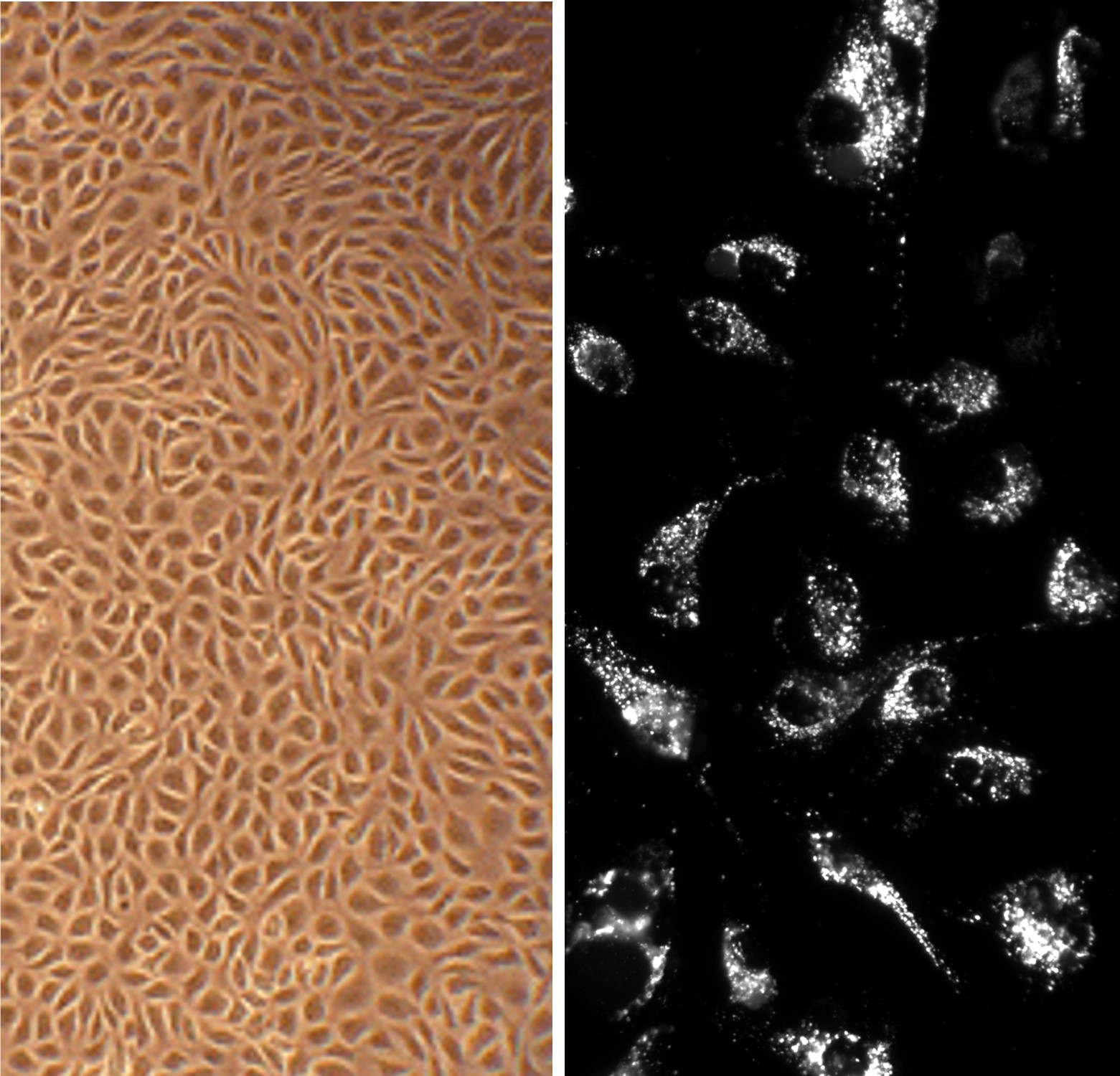
Bovine Aortic Endothelial Cell (BAOEC) monolayer (L) & BAOEC labeled w/ DiI-Ac-LDL, the acetylated apoprotein specifically recognized & endocytosed by endothelial cells (R).
Bovine Aortic Endothelial Cells: BAOEC
Primary endothelial cells isolated from the inner endothelial lining (tunica intima) of healthy bovine aorta tissue.
Description
Bovine Aortic Endothelial Cells (BAOEC), also frequently abbreviated as BAEC in scientific literature, are primary endothelial cells isolated from the inner endothelial lining (tunica intima) of healthy bovine aorta tissue. In vitro, these primary cells typically display a classic contact-inhibited cobblestone morphology at confluence. Unlike transformed cell lines, continuous endothelial cell models, or heavily modified stem-cell–derived endothelial systems, primary bovine aortic endothelial cells are used to study more physiologically relevant endothelial signaling and are typically responsive to vascular mechanobiological cues such as fluid shear stress.
To preserve a stable, non-activated phenotype, BAOEC cultures require specialized endothelial cell growth medium supplemented with optimized concentrations of serum and endothelial mitogens, according to the vendor protocol. Quality control validation often includes verifying endothelial-specific positivity (e.g., von Willebrand factor (vWF)) and Ac-LDL uptake. Standard screening pipelines for these primary cell products include ensuring lots are free of bacteria, yeast, fungi, and mycoplasma, and they are commonly distributed as cryopreserved cell pellets or living monolayers.
In the living organism, these primary endothelial cells form a continuous macromolecular monolayer lining the lumen of the aorta, an elastic conduit artery of the systemic circulatory system. Positioned at the blood–tissue interface, the aortic endothelial layer rests on an organized extracellular matrix and sits in close physical and paracrine communication with the underlying vascular smooth muscle cells of the tunica media. Macrovascular endothelial cells are adapted to withstand pulsatile hemodynamic pressures and support high-volume blood flow.
The primary biological function of BAOEC is to act as a semi-permeable physical and immunoregulatory barrier that contributes to vascular homeostasis and to the regulation of arterial tone. In homeostatic conditions, the aortic endothelium regulates vascular tone through a balance of vasorelaxing and vasoconstricting factors, including nitric oxide and endothelin-1. The endothelium also helps maintain a generally thromboresistant surface by regulating pro- and anti-thrombotic pathways, including fibrinolysis and platelet interactions.
However, exposure to mechanical disturbances, oxidized lipids, or pro-inflammatory cytokines can contribute to endothelial dysfunction. In this context, endothelial nitric oxide synthase (eNOS) activity may be reduced, and expression of adhesion molecules such as ICAM-1 and VCAM-1 can increase. This molecular activation promotes leukocyte recruitment events such as rolling, adhesion, and transendothelial migration, which are mechanistically linked to early atherogenic processes in relevant experimental models.
In vascular biology, comparative physiology, and discovery pharmacology, BAOECs are used as a large-animal primary-cell platform to investigate shear-stress mechanotransduction and cardiovascular disease mechanisms. Researchers deploy BAOEC cultures to study differential gene expression and altered protein pathways, frequently tracking protein expression by Western blot or analyzing intracellular signaling dynamics in collected cell lysates. For example, BAOEC has been used to evaluate vascular-protective effects of antioxidant or small-molecule interventions against oxidative stress–related phenotypes.
Furthermore, BAOECs can serve as a comparative benchmark for cross-species differences alongside human primary endothelial cells, mouse endothelial cell models, or endothelial-differentiated stem-cell systems. Pharmacologists and toxicologists use these cells in functional in vitro assays—such as scratch-wound migration, tube formation, and transwell permeability tests—to screen candidate therapeutics, evaluate anti-angiogenic agents, and explore strategies aimed at restoring endothelial integrity and function with appropriate cytotoxicity controls.
Bovine aortic endothelial cells (BAOEC) provide an excellent model system to study all aspects of cardiovascular function and disease, such as critical signaling pathways and mechanisms relevant to proper function of endothelia, including angiogenesis, permeability, flow adaptation, NO production, and diabetes-related complications. BAOEC can also be used to search for beneficial modulators and delivery systems for therapeutic use, and to design scaffolds, surfaces and materials for tissue engineering and 3D modeling.
BAOEC from Cell Applications, Inc. have been used in dozens of research publications, for example to:
- Determine that simvastatin modulates beta-adrenergic signaling in vascular wall by inhibiting cAMP accumulation in response to epinephrine in a mechanism involving downregulation of Galpha(s) translation via Akt/mTOR/eIF4/4EBP pathway
- Elucidate the PKA-a signaling in endothelial cells by showing that it directly phosphorylates FoxO1 to regulate expression of VCAM-1
- Identify CaMKKβ and LKB1 as critical determinants of simvastatin-dependent activation of AMPK- and Rac1-modulated signaling and reveal that Rac1 in turn regulates LKB1 phosphorylation
- Demonstrate that Chondromodulin-I (ChM-I), a cartilage-derived angiogenesis inhibitor, impairs the VEGF-A-induced Rac1/Cdc42 activity leading to destabilized lamellipodial extensions and decreased motility of endothelial cells
- Demonstrate that cofilin-mediated actin alignment in the direction of shear stress is required for endothelial barrier integrity and that p190RhoGAP links integrins and caveolin-1/caveolae to RhoA in a mechanotransduction cascade that participates in endothelial adaptation to flow
- Show that activation of RhoA/ROCK/p38 MAPK pathway causes increased endothelial arginase activity/expression and is the key mediator of endothelial dysfunction and decreased NO production precipitated by either oxidative stress-activated PKC, or by angiotensin II-activated Gα12/13 G proteins coupled to AT1 receptors
- Demonstrate that activation of mTOR/p70S6K by angiotensin II may contribute to impairment of insulin-stimulated vasodilation through phosphorylation of IRS-1 at Ser636/639
- Discover that testosterone induces a non-genomic membrane-initiated Ca2+ dependent signaling pathway that leads to activation of NF-kB, providing an explanation for why men are predisposed to earlier onset of atherosclerosis
- Demonstrate that increase in salt concentration, observed in hypertension patients or in individuals with high salt intake, suppresses NO synthase activity, contributing to the development of hypertension
- Show that valsartan, a selective angiotensin II type 1 receptor (AT1R) blocker, increases NO production via Src/PI3K/Akt signaling that leads to phosphorylation of NO synthase
- Determine that long-chain polyunsaturated fatty acids exert their beneficial effects on cardiovascular health via activation of B2 receptor leading to elevated expression of NO synthase
- Show that transient receptor potential vanilloid type 1 (TRPV1) activation by evodiamine or capsaicin initiates two Ca2+-dependent signaling pathways, both resulting in activation of NO synthase: one is PI3K/Akt/CaMKII activation, which leads to NO synthase phosphorylation at Ser1179 and Ser635, and the other involves PP2B-dependent dephosphorylation of PKC leading to decreased phosphorylation of NO synthase at Thr497
- Discover that LDL-induced endothelial disfunction is mediated by epigenetic upregulation of p66shc promoter increasing expression of p66shc which then stimulates expression of ICAM-1 and inhibits expression of thrombomodulin, leading to stimulated adhesion of monocytes and to plasma coagulation on the surface of endothelial cells
- Elucidate the involvement of AMPK cascade and autophagy in mediating beneficial cardiovascular effects of green tea;
- Show, along with Bovine Brain Microvascular Endothelial Cells, also from Cell Applications, Inc., that brain microvasculature is more sensitive to pathogenesis, compared to large vessel endothelia, by demonstrating that C-reactive protein (CRP), a cardiovascular risk factor, induces higher oxidative stress in the brain microvasculature due to higher local expression of the CRP-receptors CD16, CD32 and of the NAD(P)H-oxidase subunit p22phox, and that brain microvascular endothelial cells show higher sensitivity to oxidative stress generated by advanced glycation end products due to stronger VEGF expression leading to increased permeability
- Improve the efficiency of small molecule cancer therapeutics by prolonging their cytoplasm stay
- Construct an expression cassette to maximize targeted transgene expression in large vessel endothelia, develop new viral-based vectors an evaluate apoA-I, IL-10 and NO synthase potential for for atheroprotective human gene therapy, and design scaffolds, surfaces and materials for tissue engineering and 3D modeling, as well as for therapies aimed to prevent stent thrombosis
- Show that erythropoietin (EPO)-mediated activation of NO synthase involves AMPK-dependent signaling, which leads to enhanced phosphorylation of βCR and NO synthase
Details
| Tissue | Healthy, normal aorta of USDA-inspected cattle | |
|---|---|---|
| QC | No bacteria, yeast, fungi, mycoplasma | |
| Character | DiI-Ac-LDL uptake: Positive | |
| Bioassay | Attach, spread, proliferate in Growth Med | |
| Cryopreserved | 500,000 BAOEC (2nd passage) frozen in Basal Medium w/ 10% FBS, 10% DMSO | |
| Kit | Cryovial frozen BAOEC (B304-05), Growth Medium (B211-500), Subculture Rgnt Kit (090K) | |
| Proliferating | Shipped in Gr Med, 3rd psg (flasks or plates) | |
| Doublings | At least 16 | |
| Applications | Laboratory research use only (RUO). Not for human, clinical, diagnostic or veterinary use. |
Resources
FAQs
Need More Help?
Visit our comprehensive FAQ page for detailed answers to common questions.
Need More Help?
Visit our comprehensive FAQ page for detailed answers to common questions.
Primary Cell FAQs Powered by Bioz