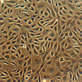
Bovine Coronary Artery Endothelial Cells: BCAEC. Isolated from blood vessel tissue.
Bovine Coronary Artery Endothelial Cells: BCAEC
Bovine Coronary Artery Endothelial Cells (BCAEC) are primary endothelial cells isolated from bovine coronary arteries.
Description
Bovine Coronary Artery Endothelial Cells (BCAEC) are primary endothelial cells isolated from bovine coronary arteries, serving as a well-established macrovascular model for investigating vascular biology, nitric oxide (NO) signaling pathways, and regional endothelial responses within the cardiac circulation.
In vivo, the endothelial cells lining the coronary arteries play a central role in maintaining cardiovascular homeostasis, regulating vascular tone, and responding to mechanical and chemical stimuli in the blood. In experimental research, BCAEC — often studied alongside human models like Human Coronary Artery Endothelial Cells (HCAEC) — can be used as a model to examine the cellular mechanisms underlying endothelial cell dysfunction, impaired vasodilation, and early processes associated with coronary artery disease and atherosclerosis. For instance, studies have utilized these cells to investigate how stress hormones such as cortisol can inhibit endothelial nitric oxide synthase (eNOS) activity and intracellular Ca2+ mobilization, as well as how specific pharmacological agents have been used to study endothelial survival, apoptotic pathways, and angiogenic potential.
Beyond cardiovascular pharmacology and disease modeling, BCAEC provide an effective in vitro platform for exploring endothelial cell proliferation, migration, and capillary-like tube formation during angiogenesis. Researchers also employ these primary cells to evaluate novel biomaterials, microstructured substrates, and mesh-like scaffolds to assess biocompatibility and cellular interactions for biomedical applications and vascular tissue engineering.
Bovine Coronary Artery Endothelial Cells (BCAEC) from CAI have been used to demonstrate that stress hormone cortisol inhibits NO synthase and Ca2+ mobilization, leading to decreased NO release by endothelial cells, resulting in increased arterial contraction and decreased blood flow. A Similar action mechanism was determined for cocaine effects on arteries. Additionally, it was found that cocaine-induced apoptosis of endothelial cells is mediated by attenuation of NO production, down-regulation of Bcl-2 and translocation of Bac to the mitochondria, followed by release of cytochrome c and activation of caspase 9 and caspase 3. BCAEC were also used to determine diabetes-related vascular dysfunction. Others employed the cells to evaluate novel mesh-like materials for their suitability for biomedical applications.

Details
| Tissue | Normal healthy bovine coronary artery | |
|---|---|---|
| QC | No bacteria, yeast, fungi, mycoplasma | |
| Character | DiI-Ac-LDL uptake: Positive | |
| Bioassay | Attach, spread, proliferate in Growth Med | |
| Cryovial | 500,000 BCAEC (2nd passage) frozen in Basal Medium w/ 10% FBS & 10% DMSO | |
| Kit | Cryovial frozen BCAEC (B300-05), Growth Med (B211-500), Subcltr Rgnt Kit (090K) | |
| Proliferating | Shipped in Gr Med, 4th psg (flasks or plates) | |
| Doublings | At least 16 | |
| Applications | Laboratory research use only (RUO). Not for human, clinical, diagnostic or veterinary use. |
Resources
FAQs
Need More Help?
Visit our comprehensive FAQ page for detailed answers to common questions.
Need More Help?
Visit our comprehensive FAQ page for detailed answers to common questions.
Primary Cell FAQs Powered by Bioz