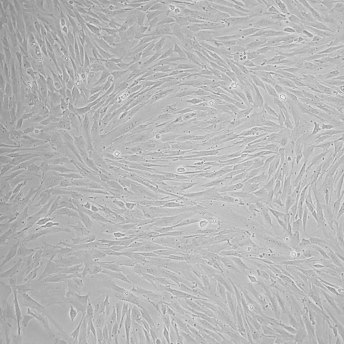
Chicken Embryonic Fibroblasts: CEF
Chicken Embryonic Fibroblasts (CEF) are primary fibroblast cells isolated from avian embryos.
Description
Chicken Embryonic Fibroblasts (CEF) — frequently referred to as CEF cells — are primary cells isolated from avian embryos that serve as a foundational model in virology, vaccine manufacturing, and cellular biology research. Characterized by an elongated, spindle-shaped morphology, these chicken embryo fibroblast cells are non-transformed primary chicken embryonic fibroblasts typically harvested from 9- to 11-day-old fertilized, specific-pathogen-free (SPF) eggs. Unlike a continuous cell line, such as the chemically immortalized chicken embryo fibroblasts known as df 1 cells, primary chicken embryo fibroblasts possess a finite lifespan in vitro. When maintained under optimized culture conditions, a high percentage of viable cells can be sustained for a limited number of passages before entering senescent phases, accurately mirroring the complex biological life stages of native chicken cells.
Within the developing chick embryo, these fibroblasts are found ubiquitously throughout the loose connective tissue and dermal layers of the body. During the window of isolation, the chicken embryo is a dense network of rapidly expanding tissue compartments where these structural cells reside embedded within a nascent extracellular matrix. To isolate them, the torso of the embryo is typically harvested, minced, and subjected to enzymatic digestion to separate the cells from the surrounding matrix. This process yields a single-cell suspension that can be plated into culture vessels, establishing a largely adherent monolayer of fibroblastic cells that represents the structural stromal framework of the developing avian organism.
In vivo, the primary function of fibroblasts is to synthesize and maintain the structural integrity of the extracellular matrix and connective tissue. These cells regulate tissue remodeling by coordinating precise windows of cell proliferation, differentiation, and programmed cell death. In vitro, the expression of their complex cellular machinery makes them an ideal host cell model for studying fundamental biological pathways. Researchers actively profile gene expression and trace changes in the cell cycle to monitor how external stimuli disrupt normal homeostasis. By mapping differentially expressed genes and tracking general proliferation dynamics, scientists can evaluate how avian cell systems respond to environmental stresses, physical injury, or intracellular pathogens.
In laboratory settings, CEF cells are a standard substrate utilized in research and certain vaccine processes. Because they are permissive to many avian viruses and specific mammalian viruses (such as Modified Vaccinia Ankara and certain influenza strains), they are frequently utilized to study viral infection and replication kinetics—though viral susceptibility varies significantly by strain and is not universal. While large-scale industrial manufacturing platforms often rely directly on embryonated eggs or established continuous mammalian and insect cell lines (such as vero cells, MDCK, or Sf9) depending on the target product, primary avian cultures remain highly relevant for poultry vaccines and specialized viral vectors. Investigators utilize advanced transfection protocols to introduce foreign genes into these host structures to study downstream protein expression and viral assembly. By indexing relevant research data under standardized mesh terms, virologists rely on infected cells in culture to analyze the exact mechanisms of viral entry, replicate life cycles, and assess the cellular defense systems of primary avian hosts.
Details
Resources
FAQs
Need More Help?
Visit our comprehensive FAQ page for detailed answers to common questions.
Need More Help?
Visit our comprehensive FAQ page for detailed answers to common questions.
Primary Cell FAQs