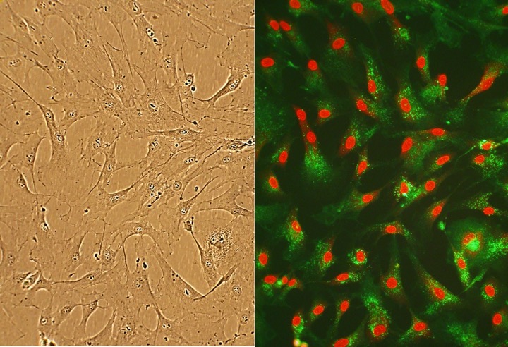
Human Cardiac Fibroblasts: HCF
Human Cardiac Fibroblasts (HCF) represent a critical fibroblast cell population in the adult heart, and are the most prevalent cell type in the heart.
Description
Human Cardiac Fibroblasts (HCF) represent a critical cell population in the adult heart, where they are classically recognized for providing the essential structural framework and maintaining the extracellular matrix (ECM) of the myocardium. Far from serving merely as passive structural support, these cells are highly dynamic coordinators of normal tissue architecture and development, as well as central drivers of pathological cardiac remodeling following injury. In a normal heart, HCF continuously monitor and regulate the homeostatic turnover of structural proteins like collagen and fibronectin, forming a supportive interstitial mesh that surrounds contractile myocytes and local capillaries.
Following myocardial injury or in response to chronic hemodynamic overload, HCF undergo active proliferation and migration, transitioning from a quiescent state into matrix-secreting myofibroblasts. This adaptive response, while initially essential for preserving mechanical stability and forming a structural scar, can become highly maladaptive if sustained. Unchecked activation results in extensive cardiac fibrosis, which increases myocardial stiffness, alters tissue compliance, and accelerates the transition toward cardiac hypertrophy and clinical heart failure.
In the laboratory, establishing a cell culture using authentic human primary cells isolated from distinct anatomical regions — such as the atrium or the ventricle — provides a high-fidelity platform to study human myocardial physiology. When maintained in an optimized fibroblast growth medium, primary HCF preserve their sensitive responsiveness to mechanical stretch, paracrine cytokines, and pharmacological agents. This establishes them as an invaluable model for modern drug discovery, enabling researchers to map the molecular mechanisms of tissue repair, screen for novel antifibrotic compounds, and investigate complex multi-lineage cellular interactions.
-
Mechanisms of Cardiac Fibrosis and Paracrine Signaling: A major focus of HCF research is dissecting how local biochemical cues stimulate phenotypic transformation. In response to mechanical stretch or ischemic stress, cardiac fibroblasts release Transforming Growth Factor-Beta (TGF-β), a potent profibrotic cytokine that plays a central role in driving fibrosis and myofibroblast differentiation. In specific experimental models, TGF-β exposure has been linked to the downregulation of thrombomodulin and subsequent prothrombotic alterations; however, these consequences are highly context-dependent. Similarly, while some studies report that the overactivation of G protein-coupled receptor kinase-2 (GRK2) can disrupt normal collagen synthesis and mimic a heart failure phenotype, investigators view this as one component of a broader, multifaceted signaling network.
-
Cardiomyocyte-Fibroblast Heterocellular Crosstalk: Maintaining coordinate contractility and rhythmicity requires intricate, reciprocal communication between distinct cell types in the heart. Investigators utilize co-culture arrays to study how HCF interact with adjacent cardiomyocyte populations and vascular endothelial cells. Specific study models suggest that heterocellular electrical coupling between myocytes and fibroblasts can be modulated by estrogen receptor agonists and may involve Ca2+-activated K+ channels. However, these electrical interactions are complex, and multiple channels alongside gap junction proteins, such as connexin43, contribute to global coupling. Additionally, literature indicates that the antimitogenic effects of estradiol on HCF growth are in part mediated by local metabolic pathways, including specific cytochrome P450 (CYP) enzymes and their downstream metabolites.
-
Ischemic Injury, Scar Formation, and Tissue Repair: Following a myocardial infarction, the rapid restoration of structural integrity to the infarcted ventricle is vital to prevent wall rupture. Researchers utilize primary HCF models to characterize the molecular pathways governing scar formation, such as exploring the precise roles of transcription factors like scleraxis and metabolic regulators like AMP-activated protein kinase alpha 1 (AMPKα1). These systems are widely deployed to evaluate novel cardioprotective agents; for example, the ATP-sensitive potassium (KATP) channel opener KMUP-3 has been reported to preserve cardiac function in some infarction models by enhancing nitric oxide (NO) synthase expression and restoring a favorable matrix metalloproteinase-9 to tissue inhibitor of metalloproteinases-1 (MMP-9/TIMP-1) balance.
-
Regenerative Medicine and Direct Reprogramming: Given the adult mammal’s highly restricted baseline capacity for cardiac regeneration, direct lineage conversion has emerged as a major frontier in treating ischemic heart disease. Investigators utilize primary HCF to optimize direct reprogramming protocols, attempting to convert structural fibroblasts into functional, beating cardiomyocyte-like cells using defined amalgams of transcription factors, microRNAs, or small molecules. By tracking the forced expression of baseline cardiogenic genes, researchers can benchmark these techniques. However, this line of conversion remains highly experimental, and both reprogramming efficiency and functional maturity remain highly protocol-dependent.
-
Fibroblast Kinetics During Heart Development: Beyond modeling adult disease states, primary cultures provide valuable comparative baselines for understanding embryonic heart development. However, researchers must account for the fact that fetal fibroblasts are phenotypically and functionally distinct from adult HCF, generally possessing a greater regenerative capacity and a less pronounced fibrotic tendency. Mapping how the fetal ECM mesh modulates cell migration, guides early chamber looping, and responds to developmental signaling networks — such as the fibroblast growth factor 2 (FGF2) pathway — helps investigators identify novel molecular targets for modulating programmed cell death and tissue expansion.
- Determine that electrical coupling between cardiomyocytes and fibroblasts is mediated by Ca2+-activated K+ channels that can be stimulated by estrogen receptor agonists
- Show that antimitogenic effects of estradiol on HCF growth are mediated by cytochromes and metabolites
- Demonstrate that in response to mechanical stretch, cardiac fibroblasts release TGF-β that downregulates trombomodulin, increases thromboembolism and induces cardiac fibroblast differentiation into myofibroblasts
- Indicate that activation of G protein-coupled receptor kinase-2 prevents normal regulation of collagen synthesis in cardiac fibroblasts mimicking heart failure phenotype
- Identify FGF2 signaling pathway as potential target for modulating apoptosis in cardiac pathology
- Investigate the roles of scleraxis and AMPKα1 in scar formation following myocardial infarction
- Show that the KATP channel opener KMUP-3 preserved cardiac function after myocardial infarction by enhancing the expression of NO synthase and restoring MMP-9/TIMP-1 balance
Details
| Tissue | Normal healthy human heart tissue | |
|---|---|---|
| QC | No bacteria, yeast, fungi, mycoplasma, virus | |
| Bioassay | Attach, spread, proliferate in Growth Med | |
| Cryovial | 500,000 HCF (1st passage) frozen in Basal Medium w/ 10% FBS, 10% DMSO | |
| Kit | Cryovial frozen HCF (306A-05a, adult atrium; 306V-05a, adult ventricle; or 306-05f fetal) , Growth Medium (316-500), Subcltr Rgnt Kit (090K) | |
| Proliferating | Shipped in Gr Med, 2nd psg (flasks or plates) | |
| Doublings | At least 8 | |
| Applications | Laboratory research use only (RUO). Not for human, clinical, diagnostic or veterinary use. |
Resources
FAQs
Need More Help?
Visit our comprehensive FAQ page for detailed answers to common questions.
Need More Help?
Visit our comprehensive FAQ page for detailed answers to common questions.
Primary Cell FAQs Powered by Bioz