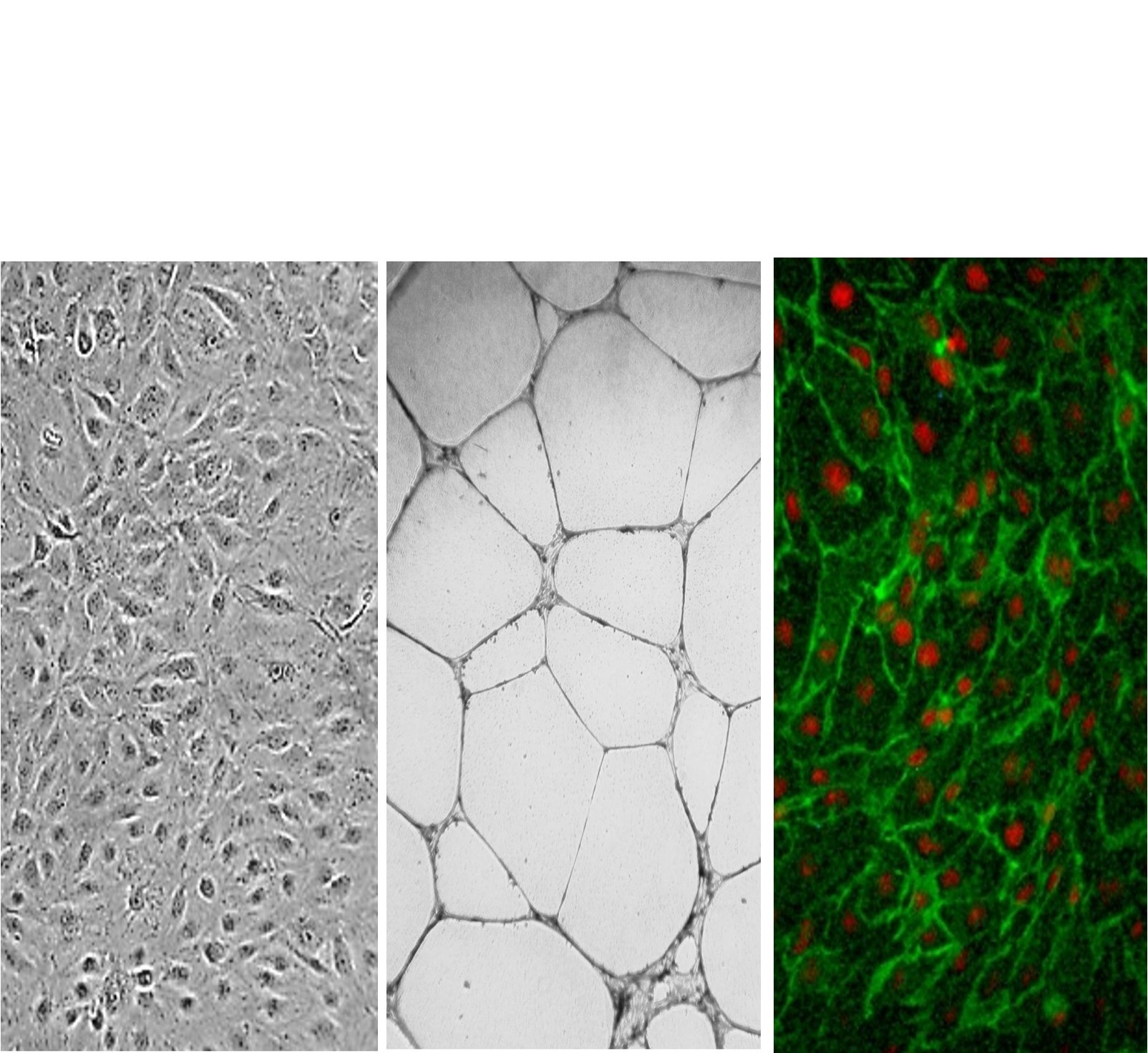
(Click to Enlarge) Cell Applications Dermal Microvascular Endothelial Cells / Human Microvascular Endothelial Cells (CADMEC / HMVEC) Mono-layer (L), vessel-like structures formed in an angiogenesis assay (M), and immunolabeled for CD31/PECAM (green). Nuclei are visualized with PI (red) (R).
Human Dermal Microvascular Endothelial Cells: CADMEC/HMVEC
Human Dermal Microvascular Endothelial Cells (CADMEC / HMVEC) are primary endothelial cells isolated from human skin capillaries.
Description
Human Dermal Microvascular Endothelial Cells (CADMEC / HMVEC) are primary endothelial cells isolated from human skin capillaries (neonatal foreskin or adult skin). In cell culture, these skin-derived microvascular cells typically display cobblestone morphology at confluence. They have been shown to express endothelial markers including CD31 (PECAM-1), vWF, and CD36, to be able to uptake DiI-Ac-LDL, and are positive for functional activity by cytokine-stimulated leukocyte adherence, though expression of some surface proteins can vary with culture conditions, passage number, confluence, and activation state.
As a primary cell population, CADMEC/HMVEC provide an in vitro tool to study microvascular signaling, distinguishing them from immortalized cell lines or transformed cancer cells that may harbor aberrant genetic mutations. To maintain their endothelial phenotype and support post-thaw recovery, they are typically cultured in specialized endothelial growth media using vendor-recommended supplements and mitogens. Quality control validation includes DiI-Ac-LDL uptake and cytokine-stimulated leukocyte adherence bioassays, along with routine screening to ensure lots are free of bacteria, yeast, fungi, and mycoplasma.
In the living organism, these cells form the inner lining of capillaries, post-capillary venules, and terminal arterioles within the dermal layers of the skin. Positioned at the blood-tissue interface, the dermal microvasculature functions in close proximity to perivascular cell types, including dermal fibroblasts, pericytes, resident immune cells, and cutaneous Schwann cells.
While macrovascular cells line large conduit blood vessels like the aorta, microvascular endothelial beds are anatomically situated to regulate localized gas exchange, nutrient delivery, and tissue fluid balance in the periphery. Because microvascular cells exhibit profound tissue-specific heterogeneity across different vascular beds, their baseline receptor profiles, junctional architecture, and metabolic constraints vary significantly. Consequently, findings established in skin-derived microvascular models like CADMEC/HMVEC cannot be universally extrapolated to other specialized environments, such as brain endothelial tight-junction models or distinct macrovascular geometries.
The primary biological function of CADMEC/HMVEC is to act as a semi-permeable physical and immunoregulatory barrier that assists in governing macromolecular flux, regulating localized tissue perfusion, and coordinating inflammatory cell trafficking. In homeostatic states, these processes are heavily influenced by endothelial nitric oxide synthase (eNOS) and nitric oxide (NO) signaling.
In response to physical trauma or pro-inflammatory cytokines — such as IL-1β, TNF-α, and IFN-γ — microvascular endothelial cells transition into an activated state. This activation drives increased production of cell adhesion molecules, such as ICAM-1 and E-selectin, which facilitate immune cell rolling, tethering, and transendothelial migration. Depending on the stimulus type, dose, and duration, activated intracellular pathways — including RhoA/ROCK and p38 MAPK or inhibition of AMPK — can promote actin cytoskeleton reorganization and increased paracellular permeability. Furthermore, under specific chronic inflammatory or fibrotic conditions, microvascular cells can undergo endothelial-to-mesenchymal transition (EndMT). This heterogeneous process can involve downregulation of endothelial markers alongside acquisition of fibroblast-like morphology and mesenchymal markers such as α-smooth muscle actin (α-SMA) or type I collagen, modulated in part by TGF-β1/Smad signaling.
In cell biology, molecular biology, and cancer research, CADMEC/HMVEC serve as a human-background model system to investigate microvascular physiology, pathological angiogenesis, and endothelial responses during wound healing. Investigators deploy these primary cells in functional in vitro assays—such as scratch-wound migration, transwell permeability flux tests, and capillary-like tube formation on extracellular matrix gels—to study downstream signaling networks and map how endothelial cell proliferation is altered by various growth factors or mechanical forces. To analyze site-specific drug sensitivities or model systemic tissue responses, researchers frequently evaluate HMVECs side-by-side with other primary human cell systems, including Human Coronary Artery Endothelial Cells (HCAEC), Human Pulmonary Artery Endothelial Cells (HPAEC), or Human Umbilical Vein Endothelial Cells (HUVEC).
In discovery pharmacology and oncology literature, these microvascular systems are utilized to evaluate cytotoxic or anti-angiogenic properties of candidate small molecules, targeted peptides, or gene delivery vehicles designed to disrupt tumor-mediated vascularization or block tumor-derived angiogenic factors. They are similarly employed as screening models to evaluate how natural products—such as specific anthocyanins or procyanidins—affect baseline oxidative stress or cytokine-induced adhesion molecule expression. Finally, in modern stem cell biology and tissue engineering, these primary cell lots serve as a reference benchmark to evaluate the transcriptomic, proteomic, and functional fidelity of endothelial cells differentiated from human pluripotent stem cells (hPSCs) or progenitor cell populations before their integration into multi-lineage 3D culturing tools or vascularized tissue constructs.
Human Dermal Microvascular Endothelial Cells (CADMEC / HMVEC) from Cell Applications, Inc. provide an excellent model system to study many aspects of endothelial function and disease, especially those related to the microvasculature and capillary systems. These cells were trademarked by Cell Applications, Inc. over 20 years ago, when they were first offered, and CADMEC™ stands for “Cell Applications’ Dermal Microvascular Endothelial Cells”. HMVEC/CADMEC have been shown to express vWF, CD36 and CD31, are able to uptake DiI-Ac-LDL and are positive for functional activity by cytokine-stimulated leukocyte adherence.
Select HMVEC lots have been additionally tested to demonstrate stimulation-dependent angiogenesis and key endothelial cell signaling pathways (phosphorylation of VEGFR2). See the Pre-Screened Endothelial Cell Product Page for more information.
CADMEC/HMVEC from Cell Applications, Inc. have been used in numerous research publications, for example to:
- Show that cytokine-activated microvascular endothelial cells upregulate expression of α2-6-linked sialic acids which are the ligands for CD22 on the surface of the B cells, increasing the adhesion between the two cell types
- Show that IL-1, TNF and IFN stimulated ICAM-1 and E-selectin production by microvascular endothelial cells and resulted in a higher leukocyte adhesion that could be prevented by dexamethasone
- Demonstrate that FGF-2, but not FGF-13, induces proliferation of microvascular endothelial cells, and that neither growth factor affected IL-6 production
- Map VEGF signaling pathway by demonstrating that c-Src phosphorylates VEGFR-2 and IQGAP1, leading to activation of b-Raf, causing endotheial cell proliferation and angiogenesis
- Demonstrate that mechanical signals promote endothelial cell proliferation via VEGFR2/Akt signaling cascade leading to inactivation of GSK3β and preventing cyclin D1 degradation
- Show that endothelial permeability caused by advanced glycation end products results from RhoA/ROCK and p38 dependent moesin phosphorylation leading to actin reorganization
- Demonstrate, along with Human Coronary Artery Endothelial Cells, also from Cell Applications, Inc., the role of AMP-activated protein kinase (AMPK) in preservation of the tight junctions, explaining the heightened vascular permeability when AMPK is inhibited due to exposure to lipopolysaccharides during sepsis
- Demonstrate that the endothelial to mesenchymal transition associated with portal venous stenosis is mediated by increased serum levels of TGF-b1 in idiopathic portal hypertension patients, which induced fibroblast-like morphology, reduction of CD34 expression, and induction of α-smooth muscle actin, COL1A1 and phospho-Smad2, and that BMP-7 was able to prevent these changes (Kitao, 2009);
- Reveal a critical role for the α7 nAChR in mediating the effects of nicotine on the endothelium
- Show that abnormal matrix composition characteristic for systemic sclerosis, leads to reduced proliferation, reduced NO-to-O2- ratio, increased apoptosis, and altered protein expression associated with endothelial to mesenchymal transition, all leading to impaired vascular function and angiogenesis
- Develop anti-tumor apoptosis-inducing peptides that selectively target angiogenic cells and a targeted gene delivery system, PEI-g-PEG-RGD, for efficient expression of soluble fragment of VEGF receptor Flt-1 (sFlt-1) gene in order to inhibit tumor-related angiogenesis
- Identify a novel tumor-derived angiogenic factor, gastrin-releasing peptide, and its small molecule inhibitor, 77427
- Evaluate, along with Human Pulmonary Artery Endothelial Cells (HPAEC) and Human Umbilical Cord Vascular Endothelial Cells (HUVEC), all obtained from Cell Applications, Inc., the cytotoxic effects of a novel pore-forming protein, proposed as an anti-tumor agent
- To confirm the antioxidant and anti-inflammatory effects of blueberry and cranberry anthocyanins and hydroxycinnamic acids against H2O2 and TNFα induced damage to microvascular endothelial cells, by demonstrating reduction in oxidative stress and lowered production of IL-8, MCP-1 and ICAM-1 and show that procyanidins isolated from cocoa inhibit the expression of ErbB2 gene, decreasing cell proliferation and angiogenesis (Kenny, 2004);
- Demonstrate secretion of adipogenic factor(s) by microvascular endothelial cells
Details
| Tissue | Normal healthy human neonatal foreskin or adult skin capillaries | |
|---|---|---|
| QC | No bacteria, yeast, fungi, mycoplasma | |
| Character | Factor VIII-related Ag, DiI-Ac-LDL uptake | |
| Bioassay | Attach, spread, proliferate in Growth Med | |
| Cryovial | 500,000 CADMEC frozen in Basal Med w/ 10% FBS, 10% DMSO | |
| Kit | Cryovial frozen CADMEC (100-05), Gr Med (112-500), Subcltr Rgnt Kit, 2 Attchmnt Factor-Coated T-25 Flasks (121-25-02), Attchmnt Fctr Soln (123-100) | |
| Proliferating | Shipped in Tsfr Med, 3rd psg (flasks or plates) | |
| Doublings | At least 16 | |
| Applications | Laboratory research use only (RUO). Not for human, clinical, diagnostic or veterinary use. |
Resources
FAQs
Need More Help?
Visit our comprehensive FAQ page for detailed answers to common questions.
Need More Help?
Visit our comprehensive FAQ page for detailed answers to common questions.
Primary Cell FAQs Powered by Bioz